Epidemiology of hepatitis D

Last updated
Worldwide prevalence of HDV among HBV carriers in 2015. Eight genotypes have been identified worldwide by comparative phylogenetic analysis. Genotype 1 is the most frequent and has variable pathogenicity, genotypes 2 and 4 are found in East Asia causing relatively mild disease. Genotype 3 is found in South America in association with severe hepatitis. Genotypes 5, 6, 7, 8 have been found only in Africa. Hep D Epidemiology Figure 1.svg
Worldwide prevalence of HDV among HBV carriers in 2015. Eight genotypes have been identified worldwide by comparative phylogenetic analysis. Genotype 1 is the most frequent and has variable pathogenicity, genotypes 2 and 4 are found in East Asia causing relatively mild disease. Genotype 3 is found in South America in association with severe hepatitis. Genotypes 5, 6, 7, 8 have been found only in Africa.

The epidemiology of hepatitis D occurs worldwide. [1] Although the figures are disputed, a recent systematic review suggests that up to 60 million individuals could be infected. [2] The major victims are the carriers of the hepatitis B surface antigen (HBsAg), who become superinfected by the HDV, and intravenous drug users who are the group at highest risk. The infection usually results in liver damage (hepatitis D); this is most often a chronic and severe hepatitis rapidly conducive to cirrhosis. [1]

Contents

Detection

Infection with the HDV is recognized by the finding of the homologous antibody (anti-HD) in serum. Testing for the viral genome (HDV RNA) is limited. In 2013, the 1st World Health Organization International Standard of HDV RNA for nucleic acid amplification techniques (NAT)-based assays was developed. [3]

The underlying HBV infection required to support the HDV is critical to determine the outcome of hepatitis D. [4] In simultaneous coinfection with the HBV, the HDV is rescued by the partner HBV with which it shares the HBsAg coat; in superinfections of HBsAg carriers, it is rescued by the foreign HBV of the carrier which provides the HBsAg coat for the assembly of the HD virion. Coinfections run an acute course; expression of the HDV is accompanied by a weak and transient antibody response and is ephemeral.

In superinfections, the chronic HBV infection and HBsAg state indefinitely sustains the replication of the HDV, resulting in a persistent anti-HD response that can be detected in any random blood sample over time; therefore, carriers of the HBsAg are the only reliable source of epidemiological information. However, HDV infections are highly pathogenic and induce the development of liver cirrhosis in approximately 70% of cases within five to ten years, with the risk of cirrhosis threefold higher in HDV-HBV co-infected than in HBV mono-infected patients. [5] As the probability of finding anti-HD throughout the clinical spectrum of HBV liver disorders increases in parallel with the severity of the liver disease. Patients with advanced HBV liver disease are the most suitable category of HBV carriers to determine the epidemiology and real health burden of HDV.

By region

Low HDV endemicity areas are North America, North Europe and Australia, where it is virtually confined to intravenous drug users and immigrants from infected areas. [6] High endemicity areas remain in the Amazon basin and low income regions of Asia and Africa; outbreaks and fulminant hepatitis D were reported in the past in the Brazilian and Peruvian Amazon, the Central African Republic, the Himalayan foothills [1] and after the year 2000, in Samara (Russia), Greenland and Mongolia. [6]

By controlling HBV infection, the implementation of hepatitis B vaccination in the industrialized world has led to a marked reduction of HDV [6] particularly in Southern Europe and Taiwan. In Italy, HDV diminished among HBV liver disorders from 24.6% in 1983 to 8% in 1997. The residual prevalence of chronic hepatitis D in HBV liver diseases in Western Europe is, as of 2010, between 4.5% and 10%, with immigrants from endemic HDV areas accounting for the larger proportion of cases. [7]

The risk of HDV has not significantly changed in recent years in countries of the world where HBV remains uncontrolled. In Asia up to 2015, the highest prevalences of chronic HDV liver disease were reported in Pakistan, Iran, Tajikistan, and Mongolia; [8] [9] a 2019 study has shown that over 80% of HBsAg cirrhosis cases in Uzbekistan are associated with HDV infection. [10] From partial and scattered information the prevalence in China, [11] and India [12] appears to be low.

In many countries of Africa, the role of hepatitis D is unknown for lack of testing. The highest rates of HDV infection were reported in sub-Saharan Africa, [13] [14] with the finding of anti-HD in over 30% and 50% of the general HBsAg population of Gabon and Cameroon, respectively, and in over 50% of the HBsAg cirrhotics in the Central African Republic (Figure 1). Lesser but consistent antibody rates (from 20% to 43%, with a mean of 24%) were reported in HBsAg liver disease in Tunisia, Mauritania, Senegal, Nigeria, Somalia and upper Egypt. [15] [16] [17] [18] Low prevalences of 2.5% and 12.7% have been reported in HBV disease carriers in Libya and Ethiopia. [19] [20] [21] [22]

Low prevalence of 0 to 8% has also been reported from Morocco, Algeria, Burkina-Faso, Benin, Mali, Sudan, South Africa and Mozambique; however, they were derived from asymptomatic HBsAg-carriers at low risk of HDV, collected at blood banks and in pregnancy clinics. [23]

Related Research Articles

<span class="mw-page-title-main">Hepatitis</span> Inflammation of the liver

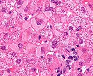
Hepatitis is inflammation of the liver tissue. Some people or animals with hepatitis have no symptoms, whereas others develop yellow discoloration of the skin and whites of the eyes (jaundice), poor appetite, vomiting, tiredness, abdominal pain, and diarrhea. Hepatitis is acute if it resolves within six months, and chronic if it lasts longer than six months. Acute hepatitis can resolve on its own, progress to chronic hepatitis, or (rarely) result in acute liver failure. Chronic hepatitis may progress to scarring of the liver (cirrhosis), liver failure, and liver cancer.

<span class="mw-page-title-main">Hepatitis C</span> Human viral infection

Hepatitis C is an infectious disease caused by the hepatitis C virus (HCV) that primarily affects the liver; it is a type of viral hepatitis. During the initial infection period, people often have mild or no symptoms. Early symptoms can include fever, dark urine, abdominal pain, and yellow tinged skin. The virus persists in the liver, becoming chronic, in about 70% of those initially infected. Early on, chronic infection typically has no symptoms. Over many years however, it often leads to liver disease and occasionally cirrhosis. In some cases, those with cirrhosis will develop serious complications such as liver failure, liver cancer, or dilated blood vessels in the esophagus and stomach.

Hepatitis D is a type of viral hepatitis caused by the hepatitis delta virus (HDV). HDV is one of five known hepatitis viruses: A, B, C, D, and E. HDV is considered to be a satellite because it can propagate only in the presence of the hepatitis B virus (HBV). Transmission of HDV can occur either via simultaneous infection with HBV (coinfection) or superimposed on chronic hepatitis B or hepatitis B carrier state (superinfection).

<span class="mw-page-title-main">Viral hepatitis</span> Liver inflammation from a viral infection

Viral hepatitis is liver inflammation due to a viral infection. It may present in acute form as a recent infection with relatively rapid onset, or in chronic form, typically progressing from a long-lasting asymptomatic condition up to a decompensated hepatic disease and hepatocellular carcinoma (HCC).

<span class="mw-page-title-main">Liver disease</span> Medical condition

Liver disease, or hepatic disease, is any of many diseases of the liver. If long-lasting it is termed chronic liver disease. Although the diseases differ in detail, liver diseases often have features in common.

<span class="mw-page-title-main">Satellite (biology)</span> Subviral agent which depends on a helper virus for its replication

A satellite is a subviral agent that depends on the coinfection of a host cell with a helper virus for its replication. Satellites can be divided into two major classes: satellite viruses and satellite nucleic acids. Satellite viruses, which are most commonly associated with plants, are also found in mammals, arthropods, and bacteria. They encode structural proteins to enclose their genetic material, which are therefore distinct from the structural proteins of their helper viruses. Satellite nucleic acids, in contrast, do not encode their own structural proteins, but instead are encapsulated by proteins encoded by their helper viruses. The genomes of satellites range upward from 359 nucleotides in length for satellite tobacco ringspot virus RNA (STobRV).

<span class="mw-page-title-main">Jade Ribbon Campaign</span> Asian Liver Center campaign

The Jade Ribbon Campaign (JRC) also known as JoinJade, was launched by the Asian Liver Center (ALC) at Stanford University in May 2001 during Asian Pacific American Heritage Month to help spread awareness internationally about hepatitis B (HBV) and liver cancer in Asian and Pacific Islander (API) communities.

In medicine, the window period for a test designed to detect a specific disease is the time between first infection and when the test can reliably detect that infection. In antibody-based testing, the window period is dependent on the time taken for seroconversion.

Hepatitis B is endemic in China. Of the 350 million individuals worldwide infected with the hepatitis B virus (HBV), one-third reside in China. As of 2006 China has immunized 11.1 million children in its poorest provinces as part of several programs initiated by the Chinese government and as part of the Global Alliance for Vaccines and Immunization (GAVI). However, the effects of these programs have yet to reach levels of immunization that would limit the spread of hepatitis B effectively.

<span class="mw-page-title-main">HBsAg</span> Surface antigen of the hepatitis B virus

HBsAg is the surface antigen of the hepatitis B virus (HBV). Its presence in blood indicates existing hepatitis B infection.

<span class="mw-page-title-main">Hepatitis B</span> Human viral infection

Hepatitis B is an infectious disease caused by the Hepatitis B virus (HBV) that affects the liver; it is a type of viral hepatitis. It can cause both acute and chronic infection.

<i>Hepatitis B virus</i> Species of the genus Orthohepadnavirus

Hepatitis B virus (HBV) is a partially double-stranded DNA virus, a species of the genus Orthohepadnavirus and a member of the Hepadnaviridae family of viruses. This virus causes the disease hepatitis B.

cccDNA is a special DNA structure that arises during the propagation of some viruses in the cell nucleus and may remain permanently there. It is a double-stranded DNA that originates in a linear form that is ligated by means of DNA ligase to a covalently closed ring. In most cases, transcription of viral DNA can occur from the circular form only. The cccDNA of viruses is also known as episomal DNA or occasionally as a minichromosome.

<span class="mw-page-title-main">Infectious causes of cancer</span> Pathogens as a cause of cancer

Estimates place the worldwide risk of cancers from infectious causes at 16.1%. Viral infections are risk factors for cervical cancer, 80% of liver cancers, and 15–20% of the other cancers. This proportion varies in different regions of the world from a high of 32.7% in Sub-Saharan Africa to 3.3% in Australia and New Zealand.

The transmission of hepadnaviruses between their natural hosts, humans, non-human primates, and birds, including intra-species host transmission and cross-species transmission, is a topic of study in virology.

<span class="mw-page-title-main">Hepatitis B virus precore mutant</span>

A precore mutant is a variety of hepatitis B virus that does not produce hepatitis B virus e antigen (HBeAg). These mutants are important because infections caused by these viruses are difficult to treat, and can cause infections of prolonged duration and with a higher risk of liver cirrhosis. The mutations are changes in DNA bases from guanine to adenine at base position 1896 (G1896A), and from cytosine to thymine at position 1858 (C1858T) in the precore region of the viral genome.

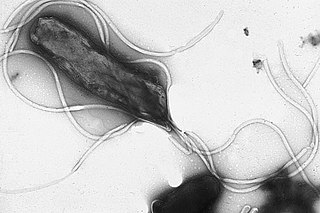
Mario Rizzetto is an Italian virologist who in 1977 first reported the Hepatitis D virus as a nuclear antigen in patients infected with HBV who had severe liver disease.

<i>Ground squirrel hepatitis virus</i> Species of virus

Ground squirrel hepatitis virus, abbreviated GSHV, is a partially double-stranded DNA virus that is closely related to human Hepatitis B virus (HBV) and Woodchuck hepatitis virus (WHV). It is a member of the family of viruses Hepadnaviridae and the genus Orthohepadnavirus. Like the other members of its family, GSHV has high degree of species and tissue specificity. It was discovered in Beechey ground squirrels, Spermophilus beecheyi, but also infects Arctic ground squirrels, Spermophilus parryi. Commonalities between GSHV and HBV include morphology, DNA polymerase activity in genome repair, cross-reacting viral antigens, and the resulting persistent infection with viral antigen in the blood (antigenemia). As a result, GSHV is used as an experimental model for HBV.

<i>Woolly monkey hepatitis B virus</i> Species of virus

The woolly monkey hepatitis B virus (WMHBV) is a viral species of the Orthohepadnavirus genus of the Hepadnaviridae family. Its natural host is the woolly monkey (Lagothrix), an inhabitant of South America categorized as a New World primate. WMHBV, like other hepatitis viruses, infects the hepatocytes, or liver cells, of its host organism. It can cause hepatitis, liver necrosis, cirrhosis, and hepatocellular carcinoma. Because nearly all species of Lagothrix are threatened or endangered, researching and developing a vaccine and/or treatment for WMHBV is important for the protection of the whole woolly monkey genus.

<span class="mw-page-title-main">Bulevirtide</span> Antiviral medication

Bulevirtide, sold under the brand name Hepcludex, is an antiviral medication for the treatment of chronic hepatitis D.

References

Open Access logo PLoS transparent.svg This article was adapted from the following source under a CC BY 4.0 license (2020) (reviewer reports): Mario Rizzetto (31 March 2020). "Epidemiology of the Hepatitis D virus" (PDF). WikiJournal of Medicine. 7 (1): 1. doi: 10.15347/WJM/2020.001.2 . ISSN   2002-4436. Wikidata   Q89093122.

  1. 1 2 3 Smedile, A; Rizzetto, M; Gerin, J (February 1994). "Advances in hepatitis D virus biology and disease". Progress in Liver Diseases. 12: 157–175. ISSN   1060-913X. OCLC   1587746. PMID   7746872.
  2. Stockdale, Alexander J; Kreuels, Benno; Henrion, Marc R Y; Giorgi, Emanuele; Kyomuhangi, Irene; Geretti, Anna Maria (2020). "Hepatitis D prevalence: problems with extrapolation to global population estimates" (PDF). Gut. 69 (2): 396–397. doi:10.1136/gutjnl-2018-317874. ISSN   0017-5749. PMID   30567743. S2CID   58554052.
  3. Chudy, Michael; Hanschmann, Kay-Martin; Bozdayi, Mithat; Kreß, Julia; Nübling, C. Micha (October 2013). "Collaborative Study to Establish a World Health Organization International Standard for Hepatitis D Virus RNA for Nucleic Acid Amplification Technique (NAT)-Based Assays" (PDF). who.int. World Health Organization . Retrieved January 2, 2020.
  4. Farci, Patrizia (2003). "Delta hepatitis: An update". Journal of Hepatology. 39: 212–219. doi:10.1016/S0168-8278(03)00331-3. PMID   14708706.
  5. Fattovich, G.; Giustina, G.; Christensen, E.; Pantalena, M.; Zagni, I.; Realdi, G.; Schalm, S. W. (2000). "Influence of hepatitis delta virus infection on morbidity and mortality in compensated cirrhosis type B". Gut. 46 (3): 420–426. doi:10.1136/gut.46.3.420. PMC   1727859 . PMID   10673308.
  6. 1 2 3 Rizzetto, Mario; Ciancio, Alessia (August 2012). "Epidemiology of Hepatitis D". Seminars in Liver Disease. 32 (3): 211–219. doi:10.1055/s-0032-1323626. ISSN   0272-8087. PMID   22932969. S2CID   23770062.
  7. Wedemeyer, Heiner; Manns, Michael P. (2010). "Epidemiology, pathogenesis and management of hepatitis D: Update and challenges ahead". Nature Reviews Gastroenterology & Hepatology. 7 (1): 31–40. doi:10.1038/nrgastro.2009.205. PMID   20051970. S2CID   12398362.
  8. Abbas, Zaigham (2010). "Hepatitis D: Scenario in the Asia-Pacific region". World Journal of Gastroenterology. 16 (5): 554–62. doi: 10.3748/wjg.v16.i5.554 . ISSN   1007-9327. PMC   2816266 . PMID   20128022.
  9. Alfaiate, Dulce; Dény, Paul; Durantel, David (2015). "Hepatitis delta virus: From biological and medical aspects to current and investigational therapeutic options". Antiviral Research. 122: 112–129. doi:10.1016/j.antiviral.2015.08.009. ISSN   0166-3542. PMID   26275800.
  10. Khodjaeva, Malika; Ibadullaeva, Nargiz; Khikmatullaeva, Aziza; Joldasova, Elizaveta; Ismoilov, Umed; Colombo, Massimo; Caviglia, Gian Paolo; Rizzetto, Mario; Musabaev, Erkin (2019). "The medical impact of hepatitis D virus infection in Uzbekistan". Liver International. 39 (11): 2077–2081. doi:10.1111/liv.14243. PMID   31505080. S2CID   202554415.
  11. Chen HY, Shen DT, Ji DZ, Han PC, Zhang WM, Ma JF, Chen WS, Goyal H, Pan S, Xu HG (March 2019). "Prevalence and burden of hepatitis D virus infection in the global population: a systematic review and meta-analysis". Gut. 68 (3): 512–521. doi:10.1136/gutjnl-2018-316601. PMID   30228220. S2CID   206969154.
  12. Jat SL, Gupta N, Kumar T, Mishra S, S A, Yadav V, Goel A, Aggarwal R (March 2015). "Prevalence of hepatitis D virus infection among hepatitis B virus-infected individuals in India". Indian J Gastroenterol. 34 (2): 164–8. doi:10.1007/s12664-015-0555-6. PMID   25902955. S2CID   20904078.
  13. Rizzetto, Mario (2019). "Hepatitis D Virus". In Wong, Robert J.; Gish, Robert G. (eds.). Clinical Epidemiology of Chronic Liver Diseases. pp. 135–148. doi:10.1007/978-3-319-94355-8_11. ISBN   978-3-319-94355-8. S2CID   239936838.
  14. Stockdale, Alexander J.; Chaponda, Mas; Beloukas, Apostolos; Phillips, Richard Odame; Matthews, Philippa C.; Papadimitropoulos, Athanasios; King, Simon; Bonnett, Laura; Geretti, Anna Maria (2017). "Prevalence of hepatitis D virus infection in sub-Saharan Africa: A systematic review and meta-analysis". The Lancet Global Health. 5 (10): e992–e1003. doi:10.1016/S2214-109X(17)30298-X. PMC   5599428 . PMID   28911765.
  15. Yacoubi L, Brichler S, Mansour W, Le Gal F, Hammami W, Sadraoui A, Ben Mami N, Msaddek A, Cheikh I, Triki H, Gordien E (November 2015). "Molecular epidemiology of hepatitis B and Delta virus strains that spread in the Mediterranean North East Coast of Tunisia". J. Clin. Virol. 72: 126–32. doi:10.1016/j.jcv.2015.10.002. PMID   26513762.
  16. Opaleye OO, Japhet OM, Adewumi OM, Omoruyi EC, Akanbi OA, Oluremi AS, Wang B, van Tong H, Velavan TP, Bock CT (April 2016). "Molecular epidemiology of hepatitis D virus circulating in Southwestern Nigeria". Virol. J. 13: 61. doi: 10.1186/s12985-016-0514-6 . PMC   4820959 . PMID   27044424.
  17. Hassan-Kadle MA, Osman MS, Ogurtsov PP (September 2018). "Epidemiology of viral hepatitis in Somalia: Systematic review and meta-analysis study". World J. Gastroenterol. 24 (34): 3927–3957. doi: 10.3748/wjg.v24.i34.3927 . PMC   6141335 . PMID   30228786.
  18. Saudy N, Sugauchi F, Tanaka Y, Suzuki S, Aal AA, Zaid MA, Agha S, Mizokami M (August 2003). "Genotypes and phylogenetic characterization of hepatitis B and delta viruses in Egypt". J. Med. Virol. 70 (4): 529–36. doi:10.1002/jmv.10427. PMID   12794714. S2CID   24724734.
  19. Elzouki AN, Bashir SM, Elahmer O, Elzouki I, Alkhattali F (December 2017). "Prevalence and risk factors of hepatitis D virus infection in patients with chronic hepatitis B infection attending the three main tertiary hospitals in Libya". Arab J Gastroenterol. 18 (4): 216–219. doi:10.1016/j.ajg.2017.11.003. PMID   29241726.
  20. Spertilli Raffaelli C, Rossetti B, Zammarchi L, Redi D, Rinaldi F, De Luca A, Montagnani F (September 2018). "Multiple chronic parasitic infections in an immunocompetent immigrant: a challenge for healthcare management". Infez Med. 26 (3): 276–279. PMID   30246773.
  21. Aberra H, Gordien E, Desalegn H, Berhe N, Medhin G, Mekasha B, Gundersen SG, Gerber A, Stene-Johansen K, Øverbø J, Johannessen A (June 2018). "Hepatitis delta virus infection in a large cohort of chronic hepatitis B patients in Ethiopia". Liver Int. 38 (6): 1000–1009. doi:10.1111/liv.13607. PMID   28980394. S2CID   24848759.
  22. Belyhun Y, Liebert UG, Maier M (September 2017). "Clade homogeneity and low rate of delta virus despite hyperendemicity of hepatitis B virus in Ethiopia". Virol. J. 14 (1): 176. doi: 10.1186/s12985-017-0844-z . PMC   5596854 . PMID   28899424.
  23. Andersson MI, Maponga TG, Ijaz S, Barnes J, Theron GB, Meredith SA, Preiser W, Tedder RS (November 2013). "The epidemiology of hepatitis B virus infection in HIV-infected and HIV-uninfected pregnant women in the Western Cape, South Africa". Vaccine. 31 (47): 5579–84. doi:10.1016/j.vaccine.2013.08.028. PMC   3898695 . PMID   23973500.