Darier's disease

Last updated
Darier's disease
Other namesDarier disease, Darier–White disease, [1] Dyskeratosis follicularis, [1] and Keratosis follicularis [2] :523 [3] :567
Specialty Medical genetics   OOjs UI icon edit-ltr-progressive.svg

Darier's disease (DD) is a rare, genetic skin disorder. It is an autosomal dominant disorder, that is, if one parent has DD, there is a 50% chance than a child will inherit DD. It was first reported by French dermatologist Ferdinand-Jean Darier in 1889.

Contents

Mild forms of the disease are the most common, consisting of skin rashes that flare up under conditions such as high humidity, high stress, or tight-fitting clothes. Short stature, combined with poorly-formed fingernails that contain vertical striations, is diagnostic even for mild forms. Symptoms usually appear in late childhood or early adulthood between the ages of about 15 and 30 years and will vary over the lifespan in an intermittent pattern of relapse (flareups) and remit.

More severe cases are characterized by dark crusty patches on the skin that are mildly greasy and that can emit a strong odor. These patches, also known as keratotic papules, keratosis follicularis, or dyskeratosis follicularis, most often appear on the arms, chest, back and legs. [4]

DD was initially studied by dermatologists. Recent research however shows DD has a whole-body effect, including cognitive (learning and intellectual) deficits, and mental health issues, particularly depression.

Diagnosis and symptoms

Rash associated with Darier's disease Darier disease rash.jpg
Rash associated with Darier's disease
Malformed, weak nails with V-shaped chips, a common sign of Darier's disease Darier disease nails.jpg
Malformed, weak nails with V-shaped chips, a common sign of Darier's disease

Diagnosis of Darier disease is often made by the appearance of the skin and nails, family history, and/or genetic testing for the mutation in the ATP2A2 gene. However, many individuals are never diagnosed because of the mildness of their symptoms. Mild cases present clinically as minor rashes (without odor) that can become exacerbated by heat, humidity, stress, and sunlight.

Clinical symptoms of the disease:[ citation needed ]

Other less common or less noticed symptoms are: [6]

Epidemiology and mental health

Worldwide prevalence of DD is estimated as between 1:30,000 and 1:100,000. Case studies have shown estimated prevalence by country to be 3.8:100,000 in Slovenia, [8] 1:36,000 in north-east England, [9] 1:30,000 in Scotland, [10] and 1:100,000 in Denmark. [11]

DD is seen in males and females equally. Symptoms typically arise between the ages of about 15 and 30, and vary over the lifetime in a relapse and remit pattern, in particular flareups that need to be managed. DD is inherited (genetic), and in particular can be traced in family groups in specific geographic localities.

Darier's disease is a non-communicable disorder, but secondary infections by bacteria and viruses can be spread to others.

DD was initially identified and studied by dermatologists (skin specialists) as a purely skin disease. Recent research however suggests DD has a whole-body effect, including cognitive and mental health issues. [12]

A study of 100 British individuals diagnosed with Darier's disease reported that affected individuals display elevated frequencies of neuropsychiatric conditions. They had high lifetime rates for mood disorders (50%), including depression (30%), bipolar disorder (4%), suicidal thoughts (31%), and suicide attempts (13%). [12]

A Swedish study of 770 individuals with DD showed a six-fold risk of being diagnosed with an intellectual disability, compared to matched Swedish population controls. [13]

A study of 76 DD patients found that 41% reported learning difficulties, notably reading difficulties, and 74% reported a family history of learning disabilities. [14] The full range of learning difficulties is not known.

Etiology and genetics

Skin changes in Darier's disease are related to a type of nutritional vitamin A deficiency that is caused by genetic mutations. The skin displays follicular dyskeratosis (degeneration of the skin in hair follicules), which reflects as hypovitaminosis A (systemic Vitamin A deficiency). [15] The skin reactions are caused by an abnormality in the desmosome-keratin filament complex leading to a breakdown in cell adhesion. [16] [5]

Mutations in a single gene, ATP2A2, are the ultimate cause for the development of Darier's disease. It is an autosomal dominant disorder, that is, if one parent has DD, there is a 50% chance than a child will inherit DD. [lower-alpha 1]

Subtypes of Darier's disease

Linear Darier's disease Darier's disease.jpg
Linear Darier's disease

Subtypes of DD have been preliminarily suggested. A large number of mutant alleles of ATP2A2 have been identified in association with Darier's Disease. One study of 19 families and 6 sporadic cases found 24 specific, novel mutations associated with DD symptoms. This study reported a loose, imperfect correlation between the severity of ATP2A2 mutations with the severity of the condition. Significant variability in disease severity between members of the same family carrying the same mutation was also reported by this study, suggesting that genetic modifiers contribute to the phenotypic penetrance of certain mutations. [20]

One subtype is linear Darier's disease. These cases result from somatic mutations to ATP2A2 in epidermal stem cells. Such individuals display phenotypic mosaicism, where the Darier's phenotype only affects the subset of epidermal tissue arising from the mutated progenitor cell.  Somatic mutations are not inherited by the offspring of such individuals. [21]

Treatment

Two recent reviews of the medical literature have evaluated treatment strategies for DD. Management and treatment of Darier disease depends on the severity of the presented clinical symptoms. Mild symptoms are often treated with moisturising creams, and more severe symptoms with topical and oral retinol or other medications (oral medications having higher strength than topical equivalents), and medical procedures. [22] [23]

In many mild cases, DD can be managed by avoiding excessive perspiration and non-breathable and abrasive clothing (producing contact dermatitis), washing with salty water, and gentle abrasion with a gauze pad. This is supplemented by moisturising lotions and topical sunscreens. Most patients with Darier's disease can live normal healthy lives.

In more severe cases of DD, application of topical and oral medications, particularly retinoids, is prescribed. Hospitalisation may be required for seriously affected individuals who display frequent relapse and remit patterns and severe infections.

Rapid resolution of rash symptoms can be complicated by the increased vulnerability of affected skin surfaces to secondary bacterial or viral infections. Bacterial overgrowth can produce an odour. The main bacteria is epidermal Staphylococcus aureus. The main viruses are human papillomavirus (HPV) and herpes simplex virus (HSV). Infections are treated with antibiotics (for bacteria) and antiviral medications (for viruses).[ citation needed ]

Treatments that have evidence-based support (though not for all persons treated) can be classified into a number of groups. [22] Because DD is a product of systemic Vitamin A deficiency, retinoids, chemical compounds (molecules) that are forms of (or related to) Vitamin A, are often recommended. Vitamin A acid compounds are often preferred as being less toxic than Vitamin A itself. [15]

1. Topical medications: Retinoids (Adapalene, Tretinoin, Isotretinoin, Tazarotene gel). Topical retinoids help in the reduction of hyperkeratosis. They work by causing the skin cells in the top layers to die and be shed off.

2. Other topical medications.

3. Oral medications: Retinoids (Acitretin, Isotretinoin). If symptoms are severe, oral retinoids have been proven to be very effective. However, there can be many adverse (and sometimes serious) side-effects associated with prolonged use. [22]

4. Medical Procedures: Surgical excision and dermabrasion, laser procedures, radiation procedures (grenz-ray, X-ray, radiology).

Support groups

Further information on and advocacy work for Darier's disease are provided by support groups.

History

Darier's disease was first described by the French dermatologist Ferdinand-Jean Darier in the journal Annales de dermatologie et de syphilographie. [26] Darier was a well-regarded dermatologist of the time who was the head of the medical department at the Hôpital Saint-Louis. Darier was an early proponent of histopathology, or the examination of samples of diseased flesh under a microscope to determine the cause of illnesses. Using this technique, he was able to uncover the origins of Darier's disease and a host of others that also bear his name. [27]

James Clarke White, a dermatologist at Harvard Medical School, independently characterized and published his observations on this dermatological disorder in the same year as Darier (1889), which is why DD is sometimes referred to as Darier-White disease. [28]

Court case

In Singapore, a man escaped the death penalty for murder as a result of Darier's disease. Ong Teng Siew, a Malaysian chicken slaughterer aged 27, was charged with murdering an 82-year-old opium addict Ng Gee Seh in December 1995. [29] [30] Ong was sentenced to death in August 1996 after the trial court found him guilty of murder, [31] and while he was appealing against his conviction, Ong was hospitalized in September 1996 for an outbreak of Darier's disease, which had previously went undiagnosed. After his lawyer discovered that the disease had a causal link to psychiatric disorders, this new evidence enabled Ong to successfully apply for a re-trial. [32] The court accepted the new evidence and that Ong was suffering from diminished responsibility as a result of Darier's disease, and therefore he was found guilty of a lesser offence of manslaughter and was re-sentenced to life imprisonment. [33] [34]

See also

Notes

  1. The gene ATP2A2 encodes the SERCA2 protein, which is a calcium pump localized to the membranes of the endoplasmic reticulum (ER) in nearly all cells and the sarcoplasmic reticulum (SR) in muscle cells. The ER is where protein processing and transport begins for proteins targeted for secretion. The SR is a specialized form of ER found in muscle cells that sequesters calcium, the regulated efflux of which into the cytosol stimulates muscle fiber contraction. Calcium acts as a second messenger in many cellular signal transduction pathways. SERCA2 is required for Ca2+ signaling in cells by removing nearly all Ca2+ ions from the cytoplasm and storing them in the ER/SR compartments. [17] [18] [16] [19]
    Darier's disease has an autosomal dominant pattern of inheritance. Autosomal dominant - en.svg
    Darier's disease has an autosomal dominant pattern of inheritance.
    The mutation is inherited in an autosomal dominant pattern. This means that only one allele needs to be mutated in order to express the trait. This also means that someone who is born to one parent with DD has a 50% chance of inheriting the mutant allele and having the disease. Loss-of-function mutations typically display recessive inheritance while the gain-of-function or hyperactive function of proteins is characteristic of dominant mutations. The observation that only one mutated allele of the SERCA2 is sufficient to produce clinical symptoms suggests that proper "gene dosage" is necessary for maintaining Ca2+ homeostasis in cells. [18] This means that two wild type copies of ATP2A2 are needed for proper cell function, which provides a logical basis for dominant phenotypes arising from loss-of-function alleles. Most ATP2A2 mutations are haploinsufficiency mutations, which means that only having only one functional copy of the functional gene results in a reduced level of protein expression that is not sufficient for wild type function for making enough of the coded protein for the cell to function properly. However, there is significant variability in disease severity in how the mutations are expressed even within families that have the same mutation. It is currently unclear in the current research why a reduction in SERCA2 expression/activity causes clinical symptoms restricted to the epidermis. One hypothesis is that other cell types express additional "back-up" Ca2+ pumps that can compensate for the reduced function or expression of the SERCA2 protein, while skin cells rely exclusively on the SERCA2 gene for calcium sequestration, meaning only they are affected by its reduction in expression. [16]

Related Research Articles

<span class="mw-page-title-main">Acne</span> Skin condition characterized by pimples

Acne also known as acne vulgaris, is a long-term skin condition that occurs when dead skin cells and oil from the skin clog hair follicles. Typical features of the condition include blackheads or whiteheads, pimples, oily skin, and possible scarring. It primarily affects skin with a relatively high number of oil glands, including the face, upper part of the chest, and back. The resulting appearance can lead to lack of confidence, anxiety, reduced self-esteem, and, in extreme cases, depression or thoughts of suicide.

<span class="mw-page-title-main">Psoriasis</span> Skin disease

Psoriasis is a long-lasting, noncontagious autoimmune disease characterized by patches of abnormal skin. These areas are red, pink, or purple, dry, itchy, and scaly. Psoriasis varies in severity from small localized patches to complete body coverage. Injury to the skin can trigger psoriatic skin changes at that spot, which is known as the Koebner phenomenon.

<span class="mw-page-title-main">Harlequin-type ichthyosis</span> Genetic skin disease

Harlequin-type ichthyosis is a genetic disorder that results in thickened skin over nearly the entire body at birth. The skin forms large, diamond/trapezoid/rectangle-shaped plates that are separated by deep cracks. These affect the shape of the eyelids, nose, mouth, and ears and limit movement of the arms and legs. Restricted movement of the chest can lead to breathing difficulties. These plates fall off over several weeks. Other complications can include premature birth, infection, problems with body temperature, and dehydration. The condition is the most severe form of ichthyosis, a group of genetic disorders characterised by scaly skin.

<span class="mw-page-title-main">Ichthyosis</span> Family of genetic skin disorders

Ichthyosis is a family of genetic skin disorders characterized by dry, thickened, scaly skin. The more than 20 types of ichthyosis range in severity of symptoms, outward appearance, underlying genetic cause and mode of inheritance. Ichthyosis comes from the Greek ἰχθύςichthys, literally 'fish', since dry, scaly skin is the defining feature of all forms of ichthyosis.

<span class="mw-page-title-main">Ichthyosis vulgaris</span> Skin disorder

Ichthyosis vulgaris is a skin disorder causing dry, scaly skin. It is the most common form, and one of the mildest forms, of ichthyosis, affecting around 1 in 250 people. For this reason it is known as common ichthyosis. It is usually an autosomal dominant inherited disease, although a rare non-heritable version called acquired ichthyosis exists.

<span class="mw-page-title-main">Epidermolytic hyperkeratosis</span> Medical condition

Epidermolytic ichthyosis (EI), is a severe form of dry scaly skin, that initially presents with redness, blisters, erosions, and peeling in a newborn baby. Hyperkeratosis typically develops several months later. Other symptoms include itch, painful fissures, strong body odor, and absence of sweat. Symptoms vary in severity and extent of skin involvement. The two main types are divided into one involving palms and soles and the other without.

<span class="mw-page-title-main">Retinoid</span> Group of tetraterpenes

The retinoids are a class of chemical compounds that are natural derivatives of vitamin A or are chemically related to it. Synthetic retinoids are used in medicine where they regulate skin health, immunity and bone disorders.

<span class="mw-page-title-main">Hyperpigmentation</span> Darkening of an area of skin or nails due to increased melanin

Hyperpigmentation is the darkening of an area of skin or nails caused by increased melanin.

<span class="mw-page-title-main">Keratosis pilaris</span> Skin condition characterized by small bumps caused by overproduction of keratin

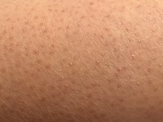
Keratosis pilaris is a common, autosomal-dominant, genetic condition of the skin's hair follicles characterized by the appearance of possibly itchy, small, gooseflesh-like bumps, with varying degrees of reddening or inflammation. It most often appears on the outer sides of the upper arms, thighs, face, back, and buttocks; KP can also occur on the hands, and tops of legs, sides, or any body part except glabrous (hairless) skin. Often the lesions can appear on the face, which may be mistaken for acne or folliculitis.

<span class="mw-page-title-main">Melasma</span> Medical condition

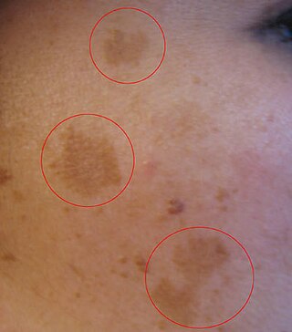
Melasma is a tan or dark skin discoloration. Melasma is thought to be caused by sun exposure, genetic predisposition, hormone changes, and skin irritation. Although it can affect anyone, it is particularly common in women, especially pregnant women and those who are taking oral or patch contraceptives or hormone replacement therapy medications.

<span class="mw-page-title-main">Meleda disease</span> Medical condition

Meleda disease (MDM) or "mal de Meleda", also called Mljet disease, keratosis palmoplantaris and transgradiens of Siemens, is an extremely rare autosomal recessive congenital skin disorder in which dry, thick patches of skin develop on the soles of the hands and feet, a condition known as palmoplantar hyperkeratosis. Meleda Disease is a skin condition which usually can be identified not long after birth. This is a genetic condition but it is very rare. The hands and feet usually are the first to show signs of the disease but the disease can advance to other parts of the body. Signs of the disease include thickening of the skin, on hands and soles of feet, which can turn red in color. There currently is no cure and treatment is limited, but Acitretin can be used in severe cases.

<span class="mw-page-title-main">Costello syndrome</span> Medical condition

Costello syndrome, also called faciocutaneoskeletal syndrome or FCS syndrome, is a rare genetic disorder that affects many parts of the body. It is characterized by delayed development and intellectual disabilities, distinctive facial features, unusually flexible joints, and loose folds of extra skin, especially on the hands and feet. Heart abnormalities are common, including a very fast heartbeat (tachycardia), structural heart defects, and overgrowth of the heart muscle. Infants with Costello syndrome may be large at birth, but grow more slowly than other children and have difficulty feeding. Later in life, people with this condition have relatively short stature and many have reduced levels of growth hormones. It is a RASopathy.

<span class="mw-page-title-main">Urbach–Wiethe disease</span> Rare recessive genetic disorder

Urbach–Wiethe disease is a very rare recessive genetic disorder, with approximately 400 reported cases since its discovery. It was first officially reported in 1929 by Erich Urbach and Camillo Wiethe, although cases may be recognized dating back as early as 1908.

Kyrle disease is identified as a form of an acquired perforating disease. Other major perforating diseases are elastosis perforans serpiginosa and reactive perforating collagenosis. Recently, however, there is a controversy on categorizing Kyrle disease with perforating dermatosis or a subtype of acquired perforating collagenosis.

<span class="mw-page-title-main">Warty dyskeratoma</span> Benign tumour

Warty dyskeratoma, also known as an Isolated dyskeratosis follicularis, is a benign epidermal proliferation with distinctive histologic findings that may mimic invasive squamous cell carcinoma and commonly manifests as an umbilicated lesion with a keratotic plug, WD have some histopathologic similarities to viral warts but it's not caused by HPV and the majority of these lesions display overall histopathologic features consistent with a follicular adnexal neoplasm. Usually limited to the head, neck, scalp or face and vulva. Lesions are generally solitary and sporadic and may be associated with a follicular unit. Oral involvement, particularly the hard palate, and genital involvement have been reported. it can also be thought of as one of the manifestations of focal acantholytic dyskeratosis, an epidermal reaction pattern that can be seen in several disorders, including Darier's disease and Grover's disease. But the main Difference between Darier disease and Warty dyskeratoma, is that Darier disease inherited dermatosis consisting of multiple keratotic papules on the face, trunk, and extremities, while WD occurs as an isolated, noninherited, single keratotic nodule mainly confined to the head and neck as mentioned earlier.

<span class="mw-page-title-main">Acrokeratosis verruciformis</span> Medical condition

Acrokeratosis verruciformis is a rare autosomal dominant disorder appearing at birth or in early childhood, characterized by skin lesions that are small, verrucous, flat papules resembling warts along with palmoplantar punctate keratoses and pits. However sporadic forms, whose less than 10 cases have been reported, presents at a later age, usually after the first decade and generally lack palmoplantar keratoses. Whether acrokeratosis verruciformis and Darier disease are related or distinct entities has been controversial, like Darier's disease, it is associated with defects in the ATP2A2 gene. however the specific mutations found in the ATP2A2 gene in acrokeratosis verruciformis have never been found in Darier's disease.

<span class="mw-page-title-main">Photoaging</span> Ultraviolet light-induced biological development over time

Photoaging or photoageing is a term used for the characteristic changes to skin induced by chronic UVA and UVB exposure. Tretinoin is the best studied retinoid in the treatment of photoaging.

Ichthyosis prematurity syndrome (IPS) is a dermatological disease with known genetic causes. This syndrome is a rare subcategory of autosomal recessive congenital ichthyosis (ARCI). It is associated with complications in the mid-trimester of a pregnancy leading to premature births. Although most prevalent in individuals of Scandinavian origin, there have also been scattered cases in people of Japanese, Italian and Indian ethnicity. This disorder is also referred to as ichthyosis congenital type IV.

<span class="mw-page-title-main">Parkes Weber syndrome</span> Medical condition

Parkes Weber syndrome (PWS) is a congenital disorder of the vascular system. It is an extremely rare condition, and its exact prevalence is unknown. It is named after British dermatologist Frederick Parkes Weber, who first described the syndrome in 1907.

<span class="mw-page-title-main">Familial disseminated comedones without dyskeratosis</span> Skin disorder from a rare gene mutation

Familial disseminated comedones without dyskeratosis (FDCWD) is a rare autosomal dominant skin disorder characterized by the presence of numerous comedones on the face, trunk, and extremities. The comedones are typically asymptomatic and do not lead to scarring. The disorder is thought to be caused by a mutation in the gene encoding the protein involucrin.

References

  1. 1 2 Rapini, Ronald P.; Bolognia, Jean L.; Jorizzo, Joseph L. (2007). Dermatology: 2-Volume Set. St. Louis, MO: Mosby. ISBN   978-1-4160-2999-1.
  2. Freedberg, et al. (2003). Fitzpatrick's Dermatology in General Medicine (6th ed.). McGraw-Hill. ISBN   0-07-138076-0.
  3. James, William; Berger, Timothy; Elston, Dirk (2005). Andrews' Diseases of the Skin: Clinical Dermatology (10th ed.). Saunders. ISBN   0-7216-2921-0.
  4. Sehgal, V. N.; Srivastava, G. (2005). "Darier's (Darier-White) disease/keratosis follicularis". International Journal of Dermatology. 44 (3): 184–192. doi:10.1111/j.1365-4632.2004.02408.x. PMID   15807723. S2CID   45303870.
  5. 1 2 3 "Darier disease NZ". DermNet. Retrieved 2020-05-07.
  6. "Darier disease | Genetic and Rare Diseases Information Center (GARD) – an NCATS Program". rarediseases.info.nih.gov. Retrieved 2020-05-08.
  7. Williams, GM; Lincoln, M (May 1, 2023). "Acrokeratosis Verruciformis of Hopf". Acrokeratosis Verruciformis of Hopf (StatPearls Internet). StatPearls. PMID   30725935.
  8. Godic A, Miljkovic J, Kansky A, Vidmar G (June 2005). "Epidemiology of Darier's Disease in Slovenia". Acta Dermatovenerol Alp Pannonica Adriat. 14 (2): 43–8. PMID   16001099.
  9. Munro CS (August 1992). "The phenotype of Darier's disease: penetrance and expressivity in adults and children". Br. J. Dermatol. 127 (2): 126–30. doi:10.1111/j.1365-2133.1992.tb08044.x. PMID   1390140. S2CID   2911858.
  10. Tavadia S, Mortimer E, Munro CS (January 2002). "Genetic epidemiology of Darier's disease: a population study in the west of Scotland". Br. J. Dermatol. 146 (1): 107–9. doi:10.1046/j.1365-2133.2002.04559.x. PMID   11841374. S2CID   42621572.
  11. Burge SM, Wilkinson JD (July 1992). "Darier-White disease: a review of the clinical features in 163 patients". J. Am. Acad. Dermatol. 27 (1): 40–50. doi:10.1016/0190-9622(92)70154-8. PMID   1619075.
  12. 1 2 Gordon-Smith K, Jones LA, Burge SM, Munro CS, Tavadia S, Craddock N (September 2010). "The neuropsychiatric phenotype in Darier disease". Br. J. Dermatol. 163 (3): 515–22. doi:10.1111/j.1365-2133.2010.09834.x. PMID   20456342. S2CID   22856369.
  13. Cederlof M, Karlsson R, et al. (July 2015). "Intellectual disability and cognitive ability in Darier disease: Swedish nation-wide study". Br. J. Dermatol. 173 (1): 155–8. doi:10.1111/bjd.13740. PMID   25704118.
  14. Dodiuk-Gad M, Lerner Z, et al. (2014). "Learning disabilities in Darier's disease patients". J. Eur. Acad. Dermat. Vener. 28 (3): 314–319. doi:10.1111/jdv.12103. PMID   23410204.
  15. 1 2 Gunther, S. (1975), "Vitamin A acid in Darier's disease.", Acta Derm Venereol Suppl (Stockh), 74: 146–151, PMID   1062882
  16. 1 2 3 Reference, Genetics Home. "Darier disease". Genetics Home Reference. Retrieved 2020-05-08.
  17. Monk, Sarah; Sakuntabhai, Anavaj; Carter, Simon A.; Bryce, Steven D.; Cox, Roger; Harrington, Louise; Levy, Elaine; Ruiz-Perez, Victor L.; Katsantoni, Eleni; Kodvawala, Ahmer; Munro, Colin S. (April 1998). "Refined Genetic Mapping of the Darier Locus to a <1-cM Region of Chromosome 12q24.1, and Construction of a Complete, High-Resolution P1 Artificial Chromosome/Bacterial Artificial Chromosome Contig of the Critical Region". The American Journal of Human Genetics. 62 (4): 890–903. doi:10.1086/301794. ISSN   0002-9297. PMC   1377034 . PMID   9529352.
  18. 1 2 Foggia, Lucie; Hovnanian, Alain (2004). "Calcium pump disorders of the skin". American Journal of Medical Genetics Part C: Seminars in Medical Genetics (in French). 131C (1): 20–31. doi: 10.1002/ajmg.c.30031 . ISSN   1552-4876. PMID   15468148. S2CID   675895.
  19. Sakuntabhai, Anavaj; Ruiz-Perez, Victor; Carter, Simon; Jacobsen, Nick; Burge, Susan; Monk, Sarah; Smith, Melanie; Munro, Colin S.; O'Donovan, Michael; Craddock, Nick; Kucherlapati, Raju (March 1999). "Mutations in ATP2A2, encoding a Ca 2+ pump, cause Darier disease". Nature Genetics. 21 (3): 271–277. doi:10.1038/6784. ISSN   1546-1718. PMID   10080178. S2CID   38684482.
  20. Ruiz-Perez, Victor L.; Carter, Simon A.; Healy, Eugene; Todd, Carole; Rees, Jonathan L.; Steijlen, Peter M.; Carmichael, Andrew J.; Lewis, Helen M.; Hohl, D.; Itin, Peter; Vahlquist, Anders (1999-09-01). "ATP2A2 Mutations in Darier's Disease: Variant Cutaneous Phenotypes Are Associated with Missense Mutations, But Neuropsychiatry Features Are Independent of Mutation Class". Human Molecular Genetics. 8 (9): 1621–1630. doi: 10.1093/hmg/8.9.1621 . ISSN   0964-6906. PMID   10441324.
  21. Sakuntabhai, Anavaj; Dhitavat, Jittima; Hovnanian, Alain; Burge, Susan (December 2000). "Mosaicism for ATP2A2 Mutations Causes Segmental Darier's Disease". Journal of Investigative Dermatology. 115 (6): 1144–1147. doi: 10.1046/j.1523-1747.2000.00182.x . ISSN   0022-202X. PMID   11121153.
  22. 1 2 3 Hanna N, Lam M, Fleming, P, Lynde CW (May–June 2022). "Therapeutic options for the treatment of Darier's disease: A comprehensive review of the literature". J. Cutan. Med. Surg. 26 (3): 280–290. doi:10.1177/12034754211058405. PMC   9125141 . PMID   34841914.
  23. Haber RM, Dib NG (Jan–Feb 2021). "Management of Darier disease: A review of the literature and update". Indian J. Dermatol. Venereol. Leprol. 87 (1): 14–21. doi: 10.25259/IJDVL_963_19 . PMID   33580925.
  24. "Dermabrasion: MedlinePlus Medical Encyclopedia". medlineplus.gov. Retrieved 2020-05-08.
  25. FIRST Skin Foundation
  26. Crissey, John Thorne; Parish, Lawrence C.; Holubar, Karl (2013). "Late nineteenth century French dermatology". Historical Atlas of Dermatology and Dermatologists. CRC Press. p. 75. ISBN   978-1-84184-864-8.
  27. Cadogan, Dr Mike (2019-03-01). "Ferdinand-Jean Darier • LITFL". Life in the Fast Lane • LITFL • Medical Blog. Retrieved 2020-05-08.
  28. "Keratosis Follicularis (Darier Disease): Background, Pathophysiology, Epidemiology". Medscape . 2020-10-01.
  29. "Was he murdered?". The New Paper. 29 December 1995.
  30. "Chicken slaughterer charged with murder". The Straits Times. 4 January 1996.
  31. "Man gets death for murdering opium addict, 82". The Straits Times. 13 August 1996.
  32. "Did man's skin disease lead him to kill addict?". The Straits Times. 24 February 1998.
  33. "Skin disease may save man's life". The Straits Times. 18 April 1998.
  34. "Skin disease saves man's neck". The Straits Times. 28 May 1998.

Sources