Reoviridae

Last updated
Sedoreoviridae
10.1371 journal.ppat.1004157.g003.ABC.png
Cryo-EM reconstruction of a rotavirus
Virus classification OOjs UI icon edit-ltr.svg
(unranked): Virus
Realm: Riboviria
Kingdom: Orthornavirae
Phylum: Duplornaviricota
Class: Resentoviricetes
Order: Reovirales
Family:Reoviridae
Subfamilies and genera

see text

Sedoreoviridae (formerly Reoviridae) is a family of double-stranded RNA viruses. Member viruses have a wide host range, including vertebrates, invertebrates, plants, protists and fungi. [1] They lack lipid envelopes and package their segmented genome within multi-layered capsids. Lack of a lipid envelope has allowed three-dimensional structures of these large complex viruses (diameter ~60–100 nm) to be obtained, revealing a structural and likely evolutionary relationship to the cystovirus family of bacteriophage. [2] There are currently 97 species in this family, divided among 15 genera in two subfamilies. [3] Reoviruses can affect the gastrointestinal system (such as rotaviruses) and respiratory tract. [4] The name "reo-" is an acronym for "respiratory enteric orphan" viruses. [5] The term "orphan virus" refers to the fact that some of these viruses have been observed not associated with any known disease. Even though viruses in the family Reoviridae have more recently been identified with various diseases, the original name is still used.

Contents

Reovirus infections occur often in humans, but most cases are mild or subclinical. Rotaviruses, however, can cause severe diarrhea and intestinal distress in children, and lab studies in mice have implicated orthoreoviruses in the expression of coeliac disease in pre-disposed individuals. [6] The virus can be readily detected in feces, and may also be recovered from pharyngeal or nasal secretions, urine, cerebrospinal fluid, and blood. Despite the ease of finding reoviruses in clinical specimens, their role in human disease or treatment is still uncertain.

Some viruses of this family, such as phytoreoviruses and oryzaviruses, infect plants. Most of the plant-infecting reoviruses are transmitted between plants by insect vectors. The viruses replicate in both the plant and the insect, generally causing disease in the plant, but little or no harm to the infected insect. [7] :148

Structure

Structure of a reovirus Viruses-07-02936-g001-A.png
Structure of a reovirus

Reoviruses are non-enveloped and have an icosahedral capsid composed of an outer (T=13) and inner (T=2) protein shell. [1] [8] Ultrastructure studies show that virion capsids are composed of two or three separate layers which depends on species type. The innermost layer (core) has T=1 icosahedral symmetry and is composed of 60 different types of structural proteins. The core contains the genome segments, each of them encode a variety enzyme structure which is required for transcription. The core is covered by capsid layer T=13 icosahedral symmetry. Reoviruses have a unique structure which is contains a glycolisated spike protein on the surface. [9]

Genome

The genomes of viruses in family Reoviridae contain 9–12 segments which are grouped into three categories corresponding to their size: L (large), M (medium) and S (small). Segments range from about 0.2 to 3 kbp and each segment encodes 1–3 proteins (10–14 proteins in total [1] ). Proteins of viruses in the family Reoviridae are denoted by the Greek character corresponding to the segment it was translated from (the L segment encodes for λ proteins, the M segment encodes for μ proteins and the S segment encodes for σ proteins). [8]

Life cycle

Life cycle of a reovirus Viruses-07-02936-g001-C.png
Life cycle of a reovirus

Viruses in the family Reoviridae have genomes consisting of segmented, double-stranded RNA (dsRNA). [4] Because of this, replication occurs exclusively in the cytoplasm, and the virus encodes several proteins which are needed for replication and conversion of the dsRNA genome into positive-sense RNAs. [10]

The virus can enter the host cell via a receptor on the cell surface. The receptor is not known but is thought to include sialic acid and junctional adhesion molecules (JAMs). [10] The virus is partially uncoated by proteases in the endolysosome, where the capsid is partially digested to allow further cell entry. The core particle then enters the cytoplasm by a yet unknown process where the genome is transcribed conservatively causing an excess of positive-sense strands, which are used as messenger RNA templates to synthesize negative-sense strands. [10]

The genome of the rotavirus is divided into 11 segments. These segments are associated with the VP1 molecule which is responsible for RNA synthesizes. In early events, the selection process occurs so that the entry of the 11 different RNA segments go in the cell. This procedure is performed by newly synthesized RNAs. This event ensures that one each of the 11 different RNA segments is received. In late events, the transcription process occurs again but this time is not capped unlike the early events. For virus different amounts of RNAs are required therefore during the translation step there is a control machinery. There are the same quantities of RNA segments but different quantities of proteins. The reason for this is that the RNA segments are not translated at the same rate. [7]

Viral particles begin to assemble in the cytoplasm 6–7 hours after infection. Translation takes place by leaky scanning, suppression of termination, and ribosomal skipping. The virus exits the host cell by monopartite non-tubule guided viral movement, cell to cell movement, and existing in occlusion bodies after cell death and remaining infectious until finding another host. [1]

Multiplicity reactivation

Multiplicity reactivation (MR) is the process by which two or more virus genomes, each containing inactivating genome damage, can interact within an infected cell to form a viable virus genome. McClain and Spendlove [11] demonstrated MR for three types of reovirus after exposure to ultraviolet irradiation. In their experiments, reovirus particles were exposed to doses of UV-light that would be lethal in single infections. However, when two or more inactivated viruses were allowed to infect individual host cells MR occurred and viable progeny were produced. As they stated, multiplicity reactivation by definition involves some type of repair. Michod et al. [12] reviewed numerous examples of MR in different viruses, and suggested that MR is a common form of sexual interaction in viruses that provides the benefit of recombinational repair of genome damages.[ citation needed ]

Taxonomy

The family Reoviridae is divided into two subfamilies [13] based on the presence of a "turret" protein on the inner capsid. [14] [15] From ICTV communications: "The name Spinareovirinae will be used to identify the subfamily containing the spiked or turreted viruses and is derived from 'reovirus' and the Latin word 'spina' as a prefix, which means spike, denoting the presence of spikes or turrets on the surface of the core particles. The term 'spiked' is an alternative to 'turreted', that was used in early research to describe the structure of the particle, particularly with the cypoviruses. The name Sedoreovirinae will be used to identify the subfamily containing the non-turreted virus genera and is derived from 'reovirus' and the Latin word 'sedo', which means smooth, denoting the absence of spikes or turrets from the core particles of these viruses, which have a relatively smooth morphology." [16]

Phylogenetic tree of family Reoviridae, dashed line divides subfamily Sedoreovirinae and Spinareovirinae 12864 2010 10194 Fig5 A HTML.png
Phylogenetic tree of family Reoviridae, dashed line divides subfamily Sedoreovirinae and Spinareovirinae

The family Reoviridae is divided into the following subfamilies and genera:

Therapeutic applications

Although reoviruses are mostly nonpathogenic in humans, these viruses have served as very productive experimental models for studies of viral pathogenesis. [17] Newborn mice are extremely sensitive to reovirus infections and have been used as the preferred experimental system for studies of reovirus pathogenesis. [2]

Reoviruses have been demonstrated to have oncolytic (cancer-killing) properties, encouraging the development of reovirus-based therapies for cancer treatment. [18] [19] [20]

Reolysin is a formulation of reovirus (Mammalian orthoreovirus serotype 3-dearing strain [21] ) that is currently in clinical trials for the treatment of various cancers, [22] including studies currently developed to investigate the role of Reolysin combined with other immunotherapies. [21]

See also

Related Research Articles

<span class="mw-page-title-main">RNA virus</span> Subclass of viruses

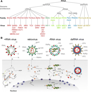
An RNA virus is a virus—other than a retrovirus—that has ribonucleic acid (RNA) as its genetic material. The nucleic acid is usually single-stranded RNA (ssRNA) but it may be double-stranded (dsRNA). Notable human diseases caused by RNA viruses include the common cold, influenza, SARS, MERS, Covid-19, Dengue Virus, hepatitis C, hepatitis E, West Nile fever, Ebola virus disease, rabies, polio, mumps, and measles.

<i>Hepadnaviridae</i> Family of viruses

Hepadnaviridae is a family of viruses. Humans, apes, and birds serve as natural hosts. There are currently 18 species in this family, divided among 5 genera. Its best-known member is hepatitis B virus. Diseases associated with this family include: liver infections, such as hepatitis, hepatocellular carcinomas, and cirrhosis. It is the sole accepted family in the order Blubervirales.

<i>Orbivirus</i> Genus of viruses

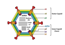
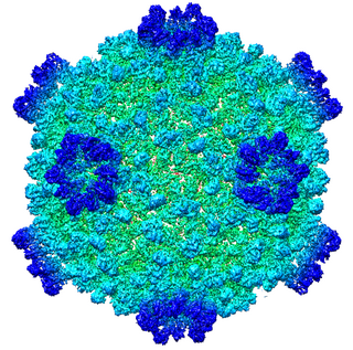
Orbivirus is a genus of double-stranded RNA viruses in the family Reoviridae and subfamily Sedoreovirinae. Unlike other reoviruses, orbiviruses are arboviruses. They can infect and replicate within a wide range of arthropod and vertebrate hosts. Orbiviruses are named after their characteristic doughnut-shaped capsomers.

<i>Herpesviridae</i> Family of DNA viruses

Herpesviridae is a large family of DNA viruses that cause infections and certain diseases in animals, including humans. The members of this family are also known as herpesviruses. The family name is derived from the Greek word ἕρπειν, referring to spreading cutaneous lesions, usually involving blisters, seen in flares of herpes simplex 1, herpes simplex 2 and herpes zoster (shingles). In 1971, the International Committee on the Taxonomy of Viruses (ICTV) established Herpesvirus as a genus with 23 viruses among four groups. As of 2020, 115 species are recognized, all but one of which are in one of the three subfamilies. Herpesviruses can cause both latent and lytic infections.

<i>Orthoreovirus</i> Genus of viruses

Orthoreovirus is a genus of viruses, in the family Reoviridae, in the subfamily Spinareovirinae. Vertebrates serve as natural hosts. There are ten species in this genus. Diseases associated with this genus include mild upper respiratory tract disease, gastroenteritis, and biliary atresia. Mammalian orthoreovirus 3 induces cell death preferentially in transformed cells and therefore displays inherent oncolytic properties.

<i>Cypovirus</i> Genus of viruses

Cypovirus, short for cytoplasmic polyhedrosis virus, is a genus of double-stranded RNA viruses in the family Reoviridae and subfamily Spinareovirinae. Cypoviruses have only been isolated from insects. Diseases associated with this genus include chronic diarrhoea and pale blue iridescence in the guts of larvae. Sixteen species are placed in this genus.

<span class="mw-page-title-main">Golden shiner virus</span> Species of virus

The golden shiner virus is an aquatic virus that infects a bait fish known as the golden shiner and to a lesser extent, aquatic animals like crustaceans and molluscs. About 6 virus species have been identified in this genus since the late 1970s. It causes death through a hemorrhagic shock. Symptoms include bleeding from the back eyes and the head. The virus is 70 nm in diameter and replicates best at 20-30 degrees Celsius. The virus has properties similar to those of the pancreatic necrosis virus. This could mean that golden shiners are more susceptible in the summer.

<span class="mw-page-title-main">Double-stranded RNA viruses</span> Type of virus according to Baltimore classification

Double-stranded RNA viruses are a polyphyletic group of viruses that have double-stranded genomes made of ribonucleic acid. The double-stranded genome is used as a template by the viral RNA-dependent RNA polymerase (RdRp) to transcribe a positive-strand RNA functioning as messenger RNA (mRNA) for the host cell's ribosomes, which translate it into viral proteins. The positive-strand RNA can also be replicated by the RdRp to create a new double-stranded viral genome.

<i>Phytoreovirus</i> Genus of viruses

Phytoreovirus is a genus of viruses, in the family Reoviridae, in the subfamily Sedoreovirinae. They are non-turreted reoviruses that are major agricultural pathogens, particularly in Asia. Oryza sativa for RDV and RGDV, dicotyledonous for WTV, and leafhoppers serve as natural hosts. There are three species in this genus. Diseases associated with this genus include: WTV: galls (tumor). RDV: dwarf disease of rice. RGDV: dwarfing, stunting, and galls.

<i>Corticovirus</i> Genus of viruses

Corticovirus is a genus of viruses in the family Corticoviridae. Corticoviruses are bacteriophages; that is, their natural hosts are bacteria. The genus contains two species. The name is derived from Latin cortex, corticis. However, prophages closely related to PM2 are abundant in the genomes of aquatic bacteria, suggesting that the ecological importance of corticoviruses might be underestimated. Bacteriophage PM2 was first described in 1968 after isolation from seawater sampled from the coast of Chile.

Avian orthoreovirus, also known as avian reovirus, is an orthoreovirus from the Reoviridae family. Infection causes arthritis and tenosynovitis in poultry. It can also cause respiratory disease.

Xi River virus (XRV) is a putative novel bat virus in the genus Orthoreovirus isolated from fruit bats in Guangdong Province in southern China. It is the first bat reovirus isolated in China.

<i>Sedoreovirinae</i> Subfamily of viruses

Sedoreovirinae is a subfamily of the Reoviridae family of viruses. Viruses in this subfamily are distinguished by the absence of a turreted protein on the inner capsid to produce a smooth surface.

Epizootic hemorrhagic disease virus, often abbreviated to EHDV, is a species of the genus Orbivirus, a member of the family Reoviridae. It is the causative agent of epizootic hemorrhagic disease, an acute, infectious, and often fatal disease of wild ruminants. In North America, the most severely affected ruminant is the white-tailed deer, although it may also infect mule deer, black-tailed deer, elk, bighorn sheep, and pronghorn antelope. It is often mistakenly referred to as “bluetongue virus” (BTV), another Orbivirus that like EHDV causes the host to develop a characteristic blue tongue due to systemic hemorrhaging and lack of oxygen in the blood. Despite showing clinical similarities, these two viruses are genetically distinct.

Mimoreovirus is a genus of viruses, in the family Reoviridae, in the subfamily Sedoreovirinae. The only isolate infects the marine photosynthetic protist Micromonas pusilla, a prasinophyte. There is only one species in this genus: Micromonas pusilla reovirus.

Triatoma virus (TrV) is a virus belonging to the insect virus family Dicistroviridae. Within this family, there are currently 3 genera and 15 species of virus. Triatoma virus belongs to the genus Cripavirus. It is non-enveloped and its genetic material is positive-sense, single-stranded RNA. The natural hosts of triatoma virus are invertebrates. TrV is a known pathogen to Triatoma infestans, the major vector of Chagas disease in Argentina which makes triatoma virus a major candidate for biological vector control as opposed to chemical insecticides. Triatoma virus was first discovered in 1984 when a survey of pathogens of triatomes was conducted in the hopes of finding potential biological control methods for T. infestans.

Umatilla virus(UMAV) is a dsRNA virus in the family Reoviridae, subfamily Sedoreovirinae, and the genus Orbivirus. This arbovirus was first isolated in 1969 in Umatilla County, Oregon in a group of Culex pipiens mosquitoes. The viral host is the Passer domesticus bird with the vectors being Culex mosquitoes.

Mammalian orthoreovirus (MRV) is a double-stranded RNA virus. It is a part of the family Reoviridae, as well as the subfamily Spinareovirinae. As seen in the name, the Mammalian Ortheoreovirus infects numerous mammalian species and vertebrates which serve as natural hosts. Some diseases that occur as a result of this virus or are associated with this virus include mild upper respiratory illness, and gastrointestinal illness. Examples of these are: upper respiratory tract syndromes, gastroenteritis, biliary atresia, obstructive hydrocephalus, jaundice, alopecia, conjunctivitis, and ‘oily hair’ associated with steatorrhea.

<i>Astroviridae</i> Family of viruses

Astroviridae is a family of non-enveloped ssRNA viruses that cause infections in different animals. The family name is derived from the Greek word astron ("star") referring to the star-like appearance of spikes projecting from the surface of these small unenveloped viruses. Astroviruses were initially identified in humans but have since been isolated from other mammals and birds. This family of viruses consists of two genera, Avastrovirus (AAstV) and Mamastrovirus (MAstV). Astroviruses most frequently cause infection of the gastrointestinal tract but in some animals they may result in encephalitis, hepatitis (avian) and nephritis (avian).

<i>Piscine orthoreovirus</i>

Piscine orthoreovirus (PRV) is a species in the genus Orthoreovirus that infects fish exclusively, PRV was first discovered in 2010 in farmed Atlantic salmon exhibiting Heart and Skeletal Muscle Inflammation (HSMI) and has been found present at higher concentration in fish with various diseases. These diseases include HSMI, jaundice syndrome, proliferative darkening syndrome and erythrocytic body inclusion syndrome. PRV is thought to mainly affect aquacultured and maricultured fish stocks, and recent research has been focused around the susceptibility of wild stock. However, whether PRV is virulent with respect to HSMI remains a topic of debate. PRV has been in the public eye mostly due to a potential linkage to farmed Atlantic Salmon exhibiting HSMI. Public concern has been raised regarding the possibility of open ocean-net farms transmitting PRV to wild salmon populations and being a factor in declining populations. PRV has not been confirmed to be pathogenic in wild salmon stocks.

References

  1. 1 2 3 4 "Viral Zone". ExPASy. Retrieved 15 June 2015.
  2. 1 2 Guglielmi, KM; Johnson, EM; Stehle, T; Dermody, TS (2006). "Attachment and Cell Entry of Mammalian Orthoreovirus". Curr Top Microbiol Immunol. Current Topics in Microbiology and Immunology. 309: 1–38. doi:10.1007/3-540-30773-7_1. ISBN   978-3-540-30772-3. PMID   16909895.
  3. "Virus Taxonomy: 2019 Release". talk.ictvonline.org. International Committee on Taxonomy of Viruses. Retrieved 11 May 2020.
  4. 1 2 Patton JT, ed. (2008). Segmented Double-stranded RNA Viruses: Structure and Molecular Biology. Caister Academic Press. ISBN   978-1-904455-21-9.
  5. Fenner, F (June 1976). "The classification and nomenclature of viruses. Summary of results of meetings of the International Committee on Taxonomy of Viruses in Madrid, September 1975". Virology. 71 (2): 371–8. doi: 10.1016/0042-6822(76)90364-0 . PMC   7131526 . PMID   820065.
  6. Bouziat, R; et al. (April 7, 2017). "Reovirus infection triggers inflammatory responses to dietary antigens and development of celiac disease". Science. 356 (6333): 44–50. Bibcode:2017Sci...356...44B. doi:10.1126/science.aah5298. PMC   5506690 . PMID   28386004.
  7. 1 2 Carter, John; Saunders, Venetia (2007). Virology: Principles and Applications. West Sussex: Wiley. ISBN   978-0-470-02386-0.
  8. 1 2 "Reoviruses". MicrobiologyBytes. Archived from the original on 2015-05-21.
  9. Payne S (2017). "Family Reoviridae". Viruses: 219–226. doi: 10.1016/B978-0-12-803109-4.00026-X . ISBN   9780128031094.
  10. 1 2 3 Barton, ES; Forrest, JC; Connolly, JL; Chappell, JD; Liu, Y; Schnell, FJ; Nusrat, A; Parkos, CA; Dermody, TS (February 9, 2001). "Junction adhesion molecule is a receptor for reovirus". Cell. 104 (3): 441–51. doi: 10.1016/S0092-8674(01)00231-8 . PMID   11239401.
  11. McClain ME, Spendlove RS (November 1966). "Multiplicity reactivation of reovirus particles after exposure to ultraviolet light". J. Bacteriol. 92 (5): 1422–9. doi:10.1128/JB.92.5.1422-1429.1966. PMC   276440 . PMID   5924273.
  12. Michod, R. E.; Bernstein, H.; Nedelcu, A. M. (2008). "Adaptive value of sex in microbial pathogens". Infection, Genetics and Evolution. 8 (3): 267–285. doi:10.1016/j.meegid.2008.01.002. PMID   18295550.
  13. Carstens, E. B. (January 2010). "Ratification vote on taxonomic proposals to the International Committee on Taxonomy of Viruses (2009)". Archives of Virology. 155 (1): 133–146. doi:10.1007/s00705-009-0547-x. PMC   7086975 . PMID   19960211.
  14. Hill C, Booth T, et al. (1999). "The structure of a cypovirus and the functional organization of dsRNA viruses". Nature Structural Biology. 6 (6): 565–9. doi:10.1038/9347. PMID   10360362. S2CID   28217302.
  15. Knipe D, Howley P, et al. (2006). Fields Virology. Philadelphia, Pa.: Wolters Kluwer, Lippincott Williams & Wilkins. p. 1855. ISBN   978-0-7817-6060-7.
  16. Attoui, Houssam; Mertens, Peter. "Template for Taxonomic Proposal to the ICTV Executive Committee To create a new SubFamily in an existing Family". International Committee on Taxonomy of Viruses. 2007.127-129V.v2.Spina-Sedoreovirinae. pp. 1–9. Archived from the original on March 5, 2010.
  17. Acheson, Nicholas H. Fundamentals of Molecular Virology. John Wiley and Sons (2011). p.234
  18. Kanai, Yuta; Kobayashi, Takeshi (29 September 2021). "FAST Proteins: Development and Use of Reverse Genetics Systems for Reoviridae Viruses". Annual Review of Virology. 8 (1): 515–536. doi: 10.1146/annurev-virology-091919-070225 . ISSN   2327-056X. PMID   34586868.
  19. Lal R, Harris D, Postel-Vinay S, de Bono J (October 2009). "Reovirus: Rationale and clinical trial update". Curr. Opin. Mol. Ther. 11 (5): 532–9. PMID   19806501.
  20. Kelland, K. (13 June 2012). "Cold virus hitches a ride to kill cancer: study". Reuters. Retrieved 17 June 2012.
  21. 1 2 Babiker, H.M.; Riaz, I.B.; Husnain, M.; Borad, M.J. (February 2017). "Oncolytic virotherapy including Rigvir and standard therapies in malignant melanoma". Oncolytic Virotherapy. Dovepress, New Zealand NLM. 6: 11–18. doi:10.2147/OV.S100072. ISSN   2253-1572. PMC   5308590 . PMID   28224120. 101629828.
  22. Thirukkumaran C, Morris DG (2009). "Oncolytic viral therapy using reovirus". Gene Therapy of Cancer. Methods in Molecular Biology. Vol. 542. pp. 607–34. doi:10.1007/978-1-59745-561-9_31. ISBN   978-1-934115-85-5. PMID   19565924.