Schiff test

Last updated
Schiff test
ClassificationColorimetric method
AnalytesAldehydes
The structure of the fuchsin dye. Fuchsin.svg
The structure of the fuchsin dye.

The Schiff test is an early organic chemistry named reaction developed by Hugo Schiff, [1] and is a relatively general chemical test for detection of many organic aldehydes that has also found use in the staining of biological tissues. [2] The Schiff reagent is the reaction product of a dye formulation such as fuchsin and sodium bisulfite; pararosaniline (which lacks an aromatic methyl group) and new fuchsin (which is uniformly mono-methylated ortho to the dye's amine functionalities) are not dye alternatives with comparable detection chemistry.

Contents

In its use as a qualitative test for aldehydes, the unknown sample is added to the decolorized Schiff reagent; when aldehyde is present a characteristic magenta color develops. Schiff-type reagents are used for various biological tissue staining methods, e.g. Feulgen stain and periodic acid-Schiff stain. Human skin also contains aldehyde functional groups in the termini of saccharides and so is stained as well.

Mechanism

Fuchsin solutions appear colored due to the visible wavelength absorbance of its central quinoid structure—see also for example viologen —but are "decolorized" upon sulfonation of the dye at its central carbon atom by sulfurous acid or its conjugate base, bisulfite. This reaction disrupts the otherwise favored delocalized extended pi-electron system and resonance in the parent molecule. [3]

The structure of the "decolorized" Schiff reagent. Schiffs Reagenz.svg
The structure of the "decolorized" Schiff reagent.

The further reaction of the Schiff reagent with aldehydes is complex with several research groups reporting multiple reaction products with model compounds. In the currently accepted mechanism, the pararosaniline and bisulfite combine to yield the "decolorized" adduct with sulfonation at the central carbon as described and shown. The free, uncharged aromatic amine groups then react with the aldehyde being tested to form two aldimine groups; these groups have also been named for their discoverer as Schiff bases (azomethines), with the usual carbinolamine (hemiaminal) intermediate being formed and dehydrated en route to the Schiff base. These electrophilic aldimine groups then react with further bisulfite, and the Ar-NH-CH(R)-SO3 product (and other resonance-stabilized species in equilibrium with the product) give rise to the magenta color of a positive test. [4] Prior formation of classical bisulfite adducts of the tested aldehyde may, when the adducts are stable, give rise to false negative tests such as in the case of testing for the aldehydic terminus of glucose. [4] Schiff's reagent on reaction with Acetaldehyde gives pink colour.

Such an imine-mediated mechanism was first proposed by Paul Rumpf (1908–1999) in 1935, [5] and experimental evidence was provided by Hardonk and van Duijn in 1964. [6] In 1980, Robins, Abrams and Pincock provided substantial NMR evidence for the mechanism, leading to its general acceptance. [7] Stoward had examined the mechanism in 1966 and, on the whole, considered this mechanism to be correct. [8]

A second, earlier mechanism continues to appear in the literature. [9] The mechanism was proposed in 1921 by the eminent German organic chemist Heinrich Wieland and his student Georg Scheuing (1895–1949). [10] [11] Bisulphite was believed to react with the available aromatic amine functional groups to form N-sulfinic acid groups, Ar-NH-SO2H, followed by reaction with aldehyde to form sulfonamides, Ar-NH-SO2CH(OH)-R. The 1980 NMR data that allowed visualization of intermediates does not support this mechanism or the sulfonamides as the chromogenic product. [7]

See also

Related Research Articles

<span class="mw-page-title-main">Gram stain</span> Investigative procedure in microbiology

Gram stain, is a method of staining used to classify bacterial species into two large groups: gram-positive bacteria and gram-negative bacteria. It may also be used to diagnose a fungal infection. The name comes from the Danish bacteriologist Hans Christian Gram, who developed the technique in 1884.

<span class="mw-page-title-main">Aldehyde</span> Organic compound containing the functional group R−CH=O

In organic chemistry, an aldehyde is an organic compound containing a functional group with the structure R−CH=O. The functional group itself can be referred to as an aldehyde but can also be classified as a formyl group. Aldehydes are a common motif in many chemicals important in technology and biology.

<span class="mw-page-title-main">Staining</span> Technique used to enhance visual contrast of specimens observed under a microscope

Staining is a technique used to enhance contrast in samples, generally at the microscopic level. Stains and dyes are frequently used in histology, in cytology, and in the medical fields of histopathology, hematology, and cytopathology that focus on the study and diagnoses of diseases at the microscopic level. Stains may be used to define biological tissues, cell populations, or organelles within individual cells.

<span class="mw-page-title-main">Sulfite</span> Oxyanion with a central atom of sulfur surrounded by 3 oxygen atoms

Sulfites or sulphites are compounds that contain the sulfite ion, SO2−
3
. The sulfite ion is the conjugate base of bisulfite. Although its acid is elusive, its salts are widely used.

<span class="mw-page-title-main">Imine</span> Organic compound or functional group containing a C=N bond

In organic chemistry, an imine is a functional group or organic compound containing a carbon–nitrogen double bond. The nitrogen atom can be attached to a hydrogen or an organic group (R). The carbon atom has two additional single bonds. Imines are common in synthetic and naturally occurring compounds and they participate in many reactions.

<span class="mw-page-title-main">Ziehl–Neelsen stain</span> Bacteriological technique

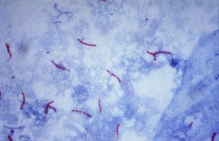
The Ziehl-Neelsen stain, also known as the acid-fast stain, is a bacteriological staining technique used in cytopathology and microbiology to identify acid-fast bacteria under microscopy, particularly members of the Mycobacterium genus. This staining method was initially introduced by Paul Ehrlich (1854–1915) and subsequently modified by the German bacteriologists Franz Ziehl (1859–1926) and Friedrich Neelsen (1854–1898) during the late 19th century.

<span class="mw-page-title-main">Fuchsine</span> Chemical compound

Fuchsine (sometimes spelled fuchsin) or rosaniline hydrochloride is a magenta dye with chemical formula C20H19N3·HCl. There are other similar chemical formulations of products sold as fuchsine, and several dozen other synonyms of this molecule.

<span class="mw-page-title-main">New fuchsine</span> Chemical compound

New fuchsine is an organic compound with the formula [(H2N(CH3)C6H3)3C]Cl. It is a green-colored solid that is used as a dye of the triarylmethane class. It is one of the four components of basic fuchsine, and one of the two that are available as single dyes. The other is pararosaniline. It is prepared by condensation of ortho-toluidine with formaldehyde. This process initially gives the benzhydrol 4,4'-bis(dimethylamino)benzhydrol, which is further condensed to give the leuco (colorless) tertiary alcohol [(H2N(CH3)C6H3)3COH, which is oxidized in acid to give the dye.
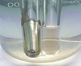
In chemistry, a chemical test is a qualitative or quantitative procedure designed to identify, quantify, or characterise a chemical compound or chemical group.

<span class="mw-page-title-main">Sulfonic acid</span> Organic compounds with the structure R−S(=O)2−OH

In organic chemistry, sulfonic acid refers to a member of the class of organosulfur compounds with the general formula R−S(=O)2−OH, where R is an organic alkyl or aryl group and the S(=O)2(OH) group a sulfonyl hydroxide. As a substituent, it is known as a sulfo group. A sulfonic acid can be thought of as sulfuric acid with one hydroxyl group replaced by an organic substituent. The parent compound is the parent sulfonic acid, HS(=O)2(OH), a tautomer of sulfurous acid, S(=O)(OH)2. Salts or esters of sulfonic acids are called sulfonates.

<span class="mw-page-title-main">Thionyl chloride</span> Inorganic compound (SOCl2)

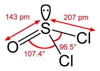
Thionyl chloride is an inorganic compound with the chemical formula SOCl2. It is a moderately volatile, colourless liquid with an unpleasant acrid odour. Thionyl chloride is primarily used as a chlorinating reagent, with approximately 45,000 tonnes per year being produced during the early 1990s, but is occasionally also used as a solvent. It is toxic, reacts with water, and is also listed under the Chemical Weapons Convention as it may be used for the production of chemical weapons.

2,4-Dinitrophenylhydrazine (2,4-DNPH or DNPH) is the organic compound C6H3(NO2)2NHNH2. DNPH is a red to orange solid. It is a substituted hydrazine. The solid is relatively sensitive to shock and friction. For this reason DNPH is usually handled as a wet powder. DNPH is a precursor to the drug Sivifene.

<span class="mw-page-title-main">Periodic acid–Schiff stain</span> Histological staining method

Periodic acid–Schiff (PAS) is a staining method used to detect polysaccharides such as glycogen, and mucosubstances such as glycoproteins, glycolipids and mucins in tissues. The reaction of periodic acid oxidizes the vicinal diols in these sugars, usually breaking up the bond between two adjacent carbons not involved in the glycosidic linkage or ring closure in the ring of the monosaccharide units that are parts of the long polysaccharides, and creating a pair of aldehydes at the two free tips of each broken monosaccharide ring. The oxidation condition has to be sufficiently regulated so as to not oxidize the aldehydes further. These aldehydes then react with the Schiff reagent to give a purple-magenta color. A suitable basic stain is often used as a counterstain.

<span class="mw-page-title-main">Sodium dithionite</span> Chemical compound

Sodium dithionite is a white crystalline powder with a sulfurous odor. Although it is stable in dry air, it decomposes in hot water and in acid solutions.

<span class="mw-page-title-main">Bisulfite</span> Chemical compound or ion

The bisulfite ion (IUPAC-recommended nomenclature: hydrogensulfite) is the ion HSO
3
. Salts containing the HSO
3
ion are also known as "sulfite lyes". Sodium bisulfite is used interchangeably with sodium metabisulfite (Na2S2O5). Sodium metabisulfite dissolves in water to give a solution of Na+HSO
3
.

<span class="mw-page-title-main">Pararosaniline</span> Chemical compound

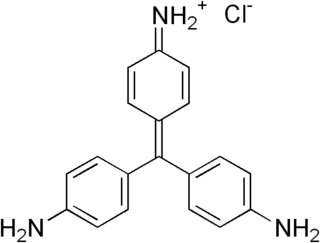
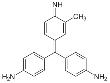
Pararosaniline, Basic Red 9, or C.I. 42500 is an organic compound with the formula [(H2NC6H4)3C]Cl. It is a magenta solid with a variety of uses as a dye. It is one of the four components of basic fuchsine. (The others are rosaniline, new fuchsine and magenta II.) It is structurally related to other triarylmethane dyes called methyl violets including crystal violet, which feature methyl groups on nitrogen.

Stephen aldehyde synthesis, a named reaction in chemistry, was invented by Henry Stephen (OBE/MBE). This reaction involves the preparation of aldehydes (R-CHO) from nitriles (R-CN) using tin(II) chloride (SnCl2), hydrochloric acid (HCl) and quenching the resulting iminium salt ([R-CH=NH2]+Cl) with water (H2O). During the synthesis, ammonium chloride is also produced.

<span class="mw-page-title-main">Acid fuchsin</span> Chemical compound

Acid fuchsin or fuchsine acid, (also called Acid Violet 19 and C.I. 42685) is an acidic magenta dye with the chemical formula C20H17N3Na2O9S3. It is a sodium sulfonate derivative of fuchsine. Acid fuchsin has wide use in histology, and is one of the dyes used in Masson's trichrome stain. This method is commonly used to stain cytoplasm and nuclei of tissue sections in the histology laboratory in order to distinguish muscle from collagen. The muscle stains red with the acid fuchsin, and the collagen is stained green or blue with Light Green SF yellowish or methyl blue. It can also be used to identify growing bacteria.

para-Dimethylaminobenzaldehyde is an organic compound containing amine and aldehyde moieties which is used in Ehrlich's reagent and Kovac's reagent to test for indoles. The carbonyl group typically reacts with the electron rich 2-position of the indole but may also react at the C-3 or N-1 positions. It may also be used for determination of hydrazine.

Verhoeff's stain, also known as Verhoeff's elastic stain (VEG) or Verhoeff–Van Gieson stain (VVG), is a staining protocol used in histology, developed by American ophthalmic surgeon and pathologist Frederick Herman Verhoeff (1874–1968) in 1908. The formulation is used to demonstrate normal or pathologic elastic fibers.

References

  1. See:
    • Schiff, Hugo (1866) "Eine neue Reihe organischer Diaminen. Zeite Abteilung." (A new series of organic diamines. Second part.), Justus Liebigs Annalen der Chemie, 140 : 92–137. [in German] From p. 132: "Das Rosanilinsulfit verbindet sich nicht direct mit den Aldehyden. Schüttelt man die rothe Lösung des krystallisirten neutralen Sulfits oder auch die nach obiger Weise bereitete leukanilinhaltige gelbe Lösung mit irgend einem flüssigen Aldehyd, so erhält man sogleich eine rothe Lösung, und diese Farbe verwandelt sich allmählich in ein je nach dem angewandten Aldehyd helleres oder dunkleres Violettblau." (Rosaniline [i.e., fuchsine] sulfite does not combine directly with aldehydes. If one shakes, with any liquid aldehyde, a red solution of the crystallized neutral sulfite [of fuchsine] or even the yellow solution which was prepared in the above-mentioned way and which contains leukaniline [i.e., fuchsine treated with aqueous sulfurous acid], then one obtains at once a red solution, and this color is transformed into a lighter or darker violet blue, depending on the aldehyde that's used.)
    • See also: Schiff, Ugo (1866) "Sopra una nova serie di basi organiche" (On a new series of organic bases), Giornale di scienze naturali ed economiche, 2 : 201–257. [in Italian] From p. 253: "Il solfito di rosanilina non si combina direttamente colle aldeidi. Quando si agita una soluzione solforosa di idrato o di acetato di rosanilina con un'aldeide qualunque, allora la soluzione gialla ássume subito un colore rosso che poco a poco si trasforma in un bel violetto più or meno intenso a secondo l'aldeide applicata." (Rosaniline sulphite does not combine directly with aldehydes. When a sulfurous solution of rosaniline hydrate or rosaniline acetate is shaken with any aldehyde, then the yellow solution immediately becomes a red color that little by little transforms into a fine violet, [which is] more or less intense according to the aldehyde [that's] used.)
    • See also: Schiff, Hugo (1867) "Dérivés de la rosaniline" (Derivatives of rosanaline [i.e., fuchsine]), Comptes rendus, 64 : 182–184. [in French] From p. 182: " … si l'on agite une solution sulfureuse diluée, soit de sulfite, soit de tout autre sel de rosaniline, avec quelques gouttes d'un aldéhyde, alors il se dégage de l'acide sulfureux, la solution se colore d'abord en rouge, puis en violet, et peu à peu il se forme un précipité constitué de petites écailles cristallines d'un violet cuivré." ( … if one shakes a dilute sulfurous solution either of the sulphite or any other salt of rosaniline [i.e., fuchsine] with a few drops of an aldehyde, then sulphurous acid is evolved; the solution is first colored red, then violet, and gradually a precipitate is formed consisting of small crystalline flakes of a coppery violet.)
  2. Histology, cell and tissue biology 5th Ed., 1983 ISBN   0-333-35406-0
  3. Juan Carlos Stockert, Alfonso Blázquez-Castro (2017). Fluorescence Microscopy in Life Sciences. Bentham Science Publishers. ISBN   978-1-68108-519-7 . Retrieved 17 December 2017.
  4. 1 2 "Schiffsche Probe" (Schiff test), University of Bayreuth, Bavaria, Germany, accessed 8 March 2013. [in German]
  5. Rumpf, P. (1935) "Recherches physico-chimiques sur la réaction colorée des aldéhydes dite “réaction de Schiff” " (Physical-chemical investigations of the color reaction of aldehydes, called the "Schiff reaction"), Annales de Chimie, 11th series, 3 : 327–442. [in French]
  6. Hardonk, M.J. & Van Duijn, P. (1964) "The mechanism of the Schiff reaction as studied with histochemical model systems," Journal of Histochemistry & Cytochemistry, 12 (10) : 748–751.
  7. 1 2 Robins, J.H., Abrams, G.D. & Pincock, J.A. (1980) "The structure of Schiff reagent aldehyde adducts and the mechanism of the Schiff reaction as determined by nuclear magnetic resonance spectroscopy," Canadian Journal of Chemistry, 58 (4) : 339–347.
  8. Stoward, P.J. (1966) "Some comments on the mechanism of the Schiff reaction," Journal of Histochemistry & Cytochemistry, 14 (9) : 681–683.
  9. Histochemistry, theoretical and applied 4th Ed. 1985 ISBN   0-443-02997-0 Note: the depiction of the sulfonic acid mechanism in this edition contains an error as the aldehyde R group is bonded to nitrogen and not to its carbon neighbor
  10. Wieland, Heinrich ; Scheuing, Georg (1921) "Die Fuchsin-schweflige Säure und ihre Farbreaktion mit Aldehyden" (Fuchsine-sulfurous acid and its colored reaction with aldehydes), Berichte der deutschen chemischen Gesellschaft, 54 (10) : 2527–2555. [in German]
  11. Puchtler, Holde; Meloan, Susan N.; Brewton, Barbara R. (1975) "On the history of basic fuchsin and aldehyde-Schiff reactions from 1862 to 1935," Histochemistry, 41 (3) : 185–194.