25-Hydroxyvitamin D 1-alpha-hydroxylase

Last updated
CYP27B1
Identifiers
Aliases CYP27B1 , CP2B, CYP1, CYP1alpha, CYP27B, P450c1, PDDR, VDD1, VDDR, VDDRI, VDR, cytochrome P450 family 27 subfamily B member 1
External IDs OMIM: 609506; MGI: 1098274; HomoloGene: 37139; GeneCards: CYP27B1; OMA:CYP27B1 - orthologs
EC number 1.14.15.18
Orthologs
SpeciesHumanMouse
Entrez
Ensembl
UniProt
RefSeq (mRNA)

NM_000785

NM_010009

RefSeq (protein)

NP_000776

NP_034139

Location (UCSC) Chr 12: 57.76 – 57.77 Mb Chr 10: 126.88 – 126.89 Mb
PubMed search [3] [4]
Wikidata
View/Edit Human View/Edit Mouse
calcidiol 1-monooxygenase
Identifiers
EC no. 1.14.15.18
CAS no. 9081-36-1
Databases
IntEnz IntEnz view
BRENDA BRENDA entry
ExPASy NiceZyme view
KEGG KEGG entry
MetaCyc metabolic pathway
PRIAM profile
PDB structures RCSB PDB PDBe PDBsum
Gene Ontology AmiGO / QuickGO
Search
PMC articles
PubMed articles
NCBI proteins

25-Hydroxyvitamin D 1-alpha-hydroxylase (VD 1A hydroxylase) also known as calcidiol 1-monooxygenase [5] or cytochrome p450 27B1 (CYP27B1) or simply 1-alpha-hydroxylase is a cytochrome P450 enzyme that in humans is encoded by the CYP27B1 gene. [6] [7] [8]

Contents

VD 1A hydroxylase is located in the proximal tubule of the kidney and a variety of other tissues, including skin (keratinocytes), immune cells, [9] and bone (osteoblasts). [10]

Reactions

The enzyme catalyzes the hydroxylation of calcifediol to calcitriol (the bioactive form of Vitamin D): [11]

calcidiol + 2 reduced adrenodoxin + 2 H+ + O2 calcitriol + 2 oxidized adrenodoxin + H2O

The enzyme is also able to oxidize ercalcidiol (25-OH D2) to ercalcitriol, secalciferol to calcitetrol, and 25-hydroxy-24-oxocalciol to (1S)-1,25-dihydroxy-24-oxocalciol. [12]

Clinical significance

Loss-of-function mutations in CYP27B1 cause Vitamin D-dependent rickets, type IA. [13]

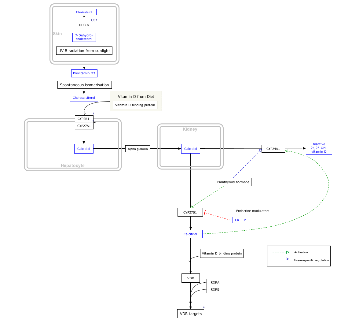
Interactive pathway map

Click on genes, proteins and metabolites below to link to respective articles. [§ 1]

[[File:
VitaminDSynthesis WP1531.png Go to articleGo to articleGo to articleGo to articlego to articleGo to articleGo to articleGo to articlego to articlego to articlego to articlego to articleGo to articleGo to articlego to articleGo to articlego to articlego to articlego to articleGo to articlego to article
[[
]]
[[
]]
[[
]]
[[
]]
[[
]]
[[
]]
[[
]]
[[
]]
[[
]]
[[
]]
[[
]]
[[
]]
[[
]]
[[
]]
[[
]]
[[
]]
[[
]]
[[
]]
[[
]]
[[
]]
[[
]]
[[
]]
VitaminDSynthesis WP1531.png Go to articleGo to articleGo to articleGo to articlego to articleGo to articleGo to articleGo to articlego to articlego to articlego to articlego to articleGo to articleGo to articlego to articleGo to articlego to articlego to articlego to articleGo to articlego to article
|alt=Vitamin D Synthesis Pathway (view / edit)]]
Vitamin D Synthesis Pathway (view / edit)
  1. The interactive pathway map can be edited at WikiPathways: "VitaminDSynthesis_WP1531".

Related Research Articles

<span class="mw-page-title-main">Cholecalciferol</span> Vitamin D3, a chemical compound

Cholecalciferol, also known as vitamin D3 or colecalciferol, is a type of vitamin D that is produced by the skin when exposed to UVB light; it is found in certain foods and can be taken as a dietary supplement.

<span class="mw-page-title-main">Calcitriol</span> Active form of vitamin D

Calcitriol is a hormone and the active form of vitamin D, normally made in the kidney. It is also known as 1,25-dihydroxycholecalciferol. It binds to and activates the vitamin D receptor in the nucleus of the cell, which then increases the expression of many genes. Calcitriol increases blood calcium mainly by increasing the uptake of calcium from the intestines.

<span class="mw-page-title-main">Vitamin D toxicity</span> Human disease

Vitamin D toxicity, or hypervitaminosis D, is the toxic state of an excess of vitamin D. The normal range for blood concentration in adults is 20 to 50 nanograms per milliliter (ng/mL).

<span class="mw-page-title-main">Fibroblast growth factor 23</span> Protein found in humans

Fibroblast growth factor 23 (FGF-23) is a protein and member of the fibroblast growth factor (FGF) family which participates in the regulation of phosphate in plasma and vitamin D metabolism. In humans it is encoded by the FGF23 gene. FGF-23 decreases reabsorption of phosphate in the kidney. Mutations in FGF23 can lead to its increased activity, resulting in autosomal dominant hypophosphatemic rickets.

<span class="mw-page-title-main">Vitamin D receptor</span> Transcription factor activated by vitamin D

The vitamin D receptor (VDR also known as the calcitriol receptor) is a member of the nuclear receptor family of transcription factors. Calcitriol (the active form of vitamin D, 1,25-(OH)2vitamin D3) binds to VDR, which then forms a heterodimer with the retinoid-X receptor. The VDR heterodimer then enters the nucleus and binds to Vitamin D responsive elements (VDRE) in genomic DNA. VDR binding results in expression or transrepression of many specific gene products. VDR is also involved in microRNA-directed post transcriptional mechanisms. In humans, the vitamin D receptor is encoded by the VDR gene located on chromosome 12q13.11.

<span class="mw-page-title-main">Aldosterone synthase</span> Protein-coding gene in the species Homo sapiens

Aldosterone synthase, also called steroid 18-hydroxylase, corticosterone 18-monooxygenase or P450C18, is a steroid hydroxylase cytochrome P450 enzyme involved in the biosynthesis of the mineralocorticoid aldosterone and other steroids. The enzyme catalyzes sequential hydroxylations of the steroid angular methyl group at C18 after initial 11β-hydroxylation. It is encoded by the CYP11B2 gene in humans.

<span class="mw-page-title-main">X-linked hypophosphatemia</span> X-linked dominant disorder that causes rickets

X-linked hypophosphatemia (XLH) is an X-linked dominant form of rickets that differs from most cases of dietary deficiency rickets in that vitamin D supplementation does not cure it. It can cause bone deformity including short stature and genu varum (bow-leggedness). It is associated with a mutation in the PHEX gene sequence (Xp.22) and subsequent inactivity of the PHEX protein. PHEX mutations lead to an elevated circulating (systemic) level of the hormone FGF23 which results in renal phosphate wasting, and local elevations of the mineralization/calcification-inhibiting protein osteopontin in the extracellular matrix of bones and teeth. An inactivating mutation in the PHEX gene results in an increase in systemic circulating FGF23, and a decrease in the enzymatic activity of the PHEX enzyme which normally removes (degrades) mineralization-inhibiting osteopontin protein; in XLH, the decreased PHEX enzyme activity leads to an accumulation of inhibitory osteopontin locally in bones and teeth to block mineralization which, along with renal phosphate wasting, both cause osteomalacia and odontomalacia.

<span class="mw-page-title-main">Calcifediol</span> Chemical compound

Calcifediol, also known as calcidiol, 25-hydroxycholecalciferol, or 25-hydroxyvitamin D3 (abbreviated 25(OH)D3), is a form of vitamin D produced in the liver by hydroxylation of vitamin D3 (cholecalciferol) by the enzyme vitamin D 25-hydroxylase. Calcifediol can be further hydroxylated by the enzyme 25(OH)D-1α-hydroxylase, primarily in the kidney, to form calcitriol (1,25-(OH)2D3), which is the active hormonal form of vitamin D.

<span class="mw-page-title-main">Calcitroic acid</span> Chemical compound

Calcitroic acid (1α-hydroxy-23-carboxy-24,25,26,27-tetranorvitamin D3) is a major metabolite of 1α,25-dihydroxyvitamin D3 (calcitriol). Around 1980, scientists first reported the isolation of calcitroic acid from the aqueous extract of radioactively treated animals' livers and intestines. Subsequent researches confirmed calcitroic acid to be a part of enterohepatic circulation. Often synthesized in the liver and kidneys, calcitroic acid is generated in the body after vitamin D is first converted into calcitriol, an intermediate in the fortification of bone through the formation and regulation of calcium in the body. These pathways managed by calcitriol are thought to be inactivated through its hydroxylation by the enzyme CYP24A1, also called calcitriol 24-hydroxylase. Specifically, It is thought to be the major route to inactivate vitamin D metabolites. The hydroxylation and oxidation reactions will yield either calcitroic acid via the C24 oxidation pathway or 1,25(OH2)D3-26,23-lactone via the C23 lactone pathway. However, the only scientifically known formation of calcitroic acid is through an oxidative reaction of the 1ɑ,25-dihydroxy vitamin D3. The positions of C24 and C23 undergo multiple oxidative reactions. Thus, causing the large and small side chains of 1ɑ,25-dihydroxy vitamin D3 to cleave off and form calcitroic acid.

<span class="mw-page-title-main">Vitamin D-binding protein</span> Mammalian protein found in Homo sapiens

Vitamin D-binding protein (DBP), also/originally known as gc-globulin, is a protein that in humans is encoded by the GC gene. DBP is genetically the oldest member of the albuminoid family and appeared early in the evolution of vertebrates.

<span class="mw-page-title-main">CYP24A1</span>

Cytochrome P450 family 24 subfamily A member 1 (abbreviated CYP24A1) is a member of the cytochrome P450 superfamily of enzymes encoded by the CYP24A1 gene. It is a mitochondrial monooxygenase which catalyzes reactions including 24-hydroxylation of calcitriol (1,25-dihydroxyvitamin D3). It has also been identified as vitamin D3 24-hydroxylase.(EC 1.14.15.16)

<span class="mw-page-title-main">Adrenal ferredoxin</span> Mammalian protein found in Homo sapiens

Adrenal ferredoxin is a protein that in humans is encoded by the FDX1 gene. In addition to the expressed gene at this chromosomal locus (11q22), there are pseudogenes located on chromosomes 20 and 21.

<span class="mw-page-title-main">CYP2R1</span> Mammalian protein found in Homo sapiens

CYP2R1 is cytochrome P450 2R1, an enzyme which is the principal vitamin D 25-hydroxylase. In humans it is encoded by the CYP2R1 gene located on chromosome 11p15.2. It is expressed in the endoplasmic reticulum in liver, where it performs the first step in the activation of vitamin D by catalyzing the formation of 25-hydroxyvitamin D.

Vitamin D<sub>5</sub> Fat soluble vitamin

Vitamin D5 (sitocalciferol) is a form of vitamin D.

<span class="mw-page-title-main">24,25-Dihydroxycholecalciferol</span> Chemical compound

24,25-Dihydroxycholecalciferol, also known as 24,25-dihydroxyvitamin D3 and (24R)-hydroxycalcidiol (abbreviated as 24(R),25-(OH)2D3), is a compound which is closely related to 1,25-dihydroxyvitamin D3, the active form of vitamin D3. Like vitamin D3 itself and calcifediol (25-hydroxyvitamin D3), it is inactive as a hormone both in vitro and in vivo. It was first identified in 1972 in the laboratory of Hector DeLuca and Michael F. Holick.

<span class="mw-page-title-main">Vitamin D deficiency</span> Human disorder

Vitamin D deficiency or hypovitaminosis D is a vitamin D level that is below normal. It most commonly occurs in people when they have inadequate exposure to sunlight, particularly sunlight with adequate ultraviolet B rays (UVB). Vitamin D deficiency can also be caused by inadequate nutritional intake of vitamin D; disorders that limit vitamin D absorption; and disorders that impair the conversion of vitamin D to active metabolites, including certain liver, kidney, and hereditary disorders. Deficiency impairs bone mineralization, leading to bone-softening diseases, such as rickets in children. It can also worsen osteomalacia and osteoporosis in adults, increasing the risk of bone fractures. Muscle weakness is also a common symptom of vitamin D deficiency, further increasing the risk of fall and bone fractures in adults. Vitamin D deficiency is associated with the development of schizophrenia.

<span class="mw-page-title-main">Alfacalcidol</span> Chemical compound

Alfacalcidol is an analogue of vitamin D used for supplementation in humans and as a poultry feed additive.

<span class="mw-page-title-main">Vitamin D</span> Group of fat-soluble secosteroids

Vitamin D is a group of fat-soluble secosteroids responsible for increasing intestinal absorption of calcium, magnesium, and phosphate, along with numerous other biological functions. In humans, the most significant compounds within this group are vitamin D3 (cholecalciferol) and vitamin D2 (ergocalciferol).

<span class="mw-page-title-main">Michael F. Holick</span> American physician–scientist

Michael F. Holick is an American adult endocrinologist, specializing in vitamin D, such as the identification of both calcidiol, the major circulating form of vitamin D, and calcitriol, the active form of vitamin D. His work has been the basis for diagnostic tests and therapies for vitamin D-related diseases. He is a professor of medicine at the Boston University Medical Center and editor-in-chief of the journal Clinical Laboratory.

Vitamin D3 24-hydroxylase (EC 1.14.15.16, CYP24A1) is an enzyme with systematic name calcitriol,NADPH:oxygen oxidoreductase (24-hydroxylating). This enzyme catalyses the following chemical reaction

References

  1. 1 2 3 GRCh38: Ensembl release 89: ENSG00000111012 Ensembl, May 2017
  2. 1 2 3 GRCm38: Ensembl release 89: ENSMUSG00000006724 Ensembl, May 2017
  3. "Human PubMed Reference:". National Center for Biotechnology Information, U.S. National Library of Medicine.
  4. "Mouse PubMed Reference:". National Center for Biotechnology Information, U.S. National Library of Medicine.
  5. "25-Hydroxyvitamin D3 1-Alpha-Hydroxylase - an overview | ScienceDirect Topics".
  6. "Entrez Gene: cytochrome P450".
  7. Takeyama K, Kitanaka S, Sato T, Kobori M, Yanagisawa J, Kato S (Sep 1997). "25-Hydroxyvitamin D3 1alpha-hydroxylase and vitamin D synthesis". Science. 277 (5333): 1827–30. doi:10.1126/science.277.5333.1827. PMID   9295274.
  8. Monkawa T, Yoshida T, Wakino S, Shinki T, Anazawa H, Deluca HF, Suda T, Hayashi M, Saruta T (Oct 1997). "Molecular cloning of cDNA and genomic DNA for human 25-hydroxyvitamin D3 1 alpha-hydroxylase". Biochemical and Biophysical Research Communications. 239 (2): 527–33. doi:10.1006/bbrc.1997.7508. PMID   9344864.
  9. Sigmundsdottir H, Pan J, Debes GF, Alt C, Habtezion A, Soler D, Butcher EC (Mar 2007). "DCs metabolize sunlight-induced vitamin D3 to 'program' T cell attraction to the epidermal chemokine CCL27" (PDF). Nature Immunology. 8 (3): 285–93. doi:10.1038/ni1433. PMID   17259988. S2CID   9540123.[ permanent dead link ]
  10. Kogawa M, Findlay DM, Anderson PH, Ormsby R, Vincent C, Morris HA, Atkins GJ (Oct 2010). "Osteoclastic metabolism of 25(OH)-vitamin D3: a potential mechanism for optimization of bone resorption". Endocrinology. 151 (10): 4613–25. doi: 10.1210/en.2010-0334 . PMID   20739402.
  11. Gray RW, Omdahl JL, Ghazarian JG, DeLuca HF (Dec 1972). "25-Hydroxycholecalciferol-1-hydroxylase. Subcellular location and properties". The Journal of Biological Chemistry. 247 (23): 7528–32. doi: 10.1016/S0021-9258(19)44557-2 . PMID   4404596.
  12. Sawada, N; Sakaki, T; Kitanaka, S; Takeyama, K; Kato, S; Inouye, K (November 1999). "Enzymatic properties of human 25-hydroxyvitamin D3 1alpha-hydroxylase coexpression with adrenodoxin and NADPH-adrenodoxin reductase in Escherichia coli". European Journal of Biochemistry. 265 (3): 950–6. doi: 10.1046/j.1432-1327.1999.00794.x . PMID   10518789.
  13. "# 264700 - VITAMIN D HYDROXYLATION-DEFICIENT RICKETS, TYPE 1A; VDDR1A". www.omim.org.

Further reading