Index of trauma and orthopaedics articles

Last updated

The Tree of Andry Andry tree.png
The Tree of Andry

Orthopedic surgery is the branch of surgery concerned with conditions involving the musculoskeletal system. Orthopedic surgeons use both surgical and nonsurgical means to treat musculoskeletal injuries, sports injuries, degenerative diseases, infections, bone tumours, and congenital limb deformities. Trauma surgery and traumatology is a sub-specialty dealing with the operative management of fractures, major trauma and the multiply-injured patient.

Contents

List excludes anatomical terminology covered in index of anatomy articles.

A

Abbreviated Injury Scale - Acetabular fracture - Acheiropodia - Achilles tendon rupture - Acromioplasty - Adamantinoma - Adhesive capsulitis of shoulder - Advanced trauma life support - Ainhum - Akin osteotomy - Albers-Schonberg disease - Albright's hereditary osteodystrophy - Allis test - ALPSA lesion - Amelia (birth defect) - American Joint Replacement Registry - Amphiarthrosis - Andersson lesion - Aneurysmal bone cyst - Ankle replacement - Anterior cruciate ligament injury - Anterior cruciate ligament reconstruction - Antley–Bixler syndrome - Apert syndrome - Apley grind test - Apley scratch test - Apprehension test - Arachnodactyly - Arm fracture - Arthralgia - Arthritis - Arthrocentesis - Arthrodesis - Arthrogram - Arthrogryposis - Arthroplasty - Arthroscopy - Arthrotomy - Articular capsule - Articular cartilage repair - Astragalectomy - Autologous chondrocyte implantation - Avascular necrosis - Avulsion fracture

B

Baastrup's sign - Baker's cyst - Baksi's prosthesis - Ballottement - Bankart lesion - Bankart's fracture - Barlow maneuver - Barré–Liéou syndrome - Barton's fracture - Baumann's angle - Beals syndrome - Bechterew's - Bennett's fracture - Bifid rib - Bimalleolar fracture - Blount's disease - Blumensaat's line - Blunt trauma - Bohler's angle - Bone cutter - Bone cyst - Bone density - Bone disease - Bone fracture - Bone fracture healing - Bone grafting - Bone healing - Bone metastases - Bone mineral - Bone pathology - Bone remodeling - Bone resorption - Bone tumor - Bone - Bosworth fracture - Bouchard's nodes - Boutonniere deformity - Boxer's fracture - Brachydactyly - British Orthopaedic Association - Brodie abscess - Broström procedure - Brown tumor - Bruck syndrome - Brunelli procedure - Bryant's traction - Buddy wrapping - Bumper fracture - Bunion - Burst fracture

C

Calcaneal fracture - Camurati–Engelmann disease - Cancellous bone - Cartilage - Cartilaginous joint - Catel–Manzke syndrome - Cenani–Lenz syndactylism - Cervical dislocation - Cervical fracture - Cervical rib - Chalkstick fracture - Chance fracture - Chandler's disease - Charnley prosthesis - Charnley retractor - Chauffeur's fracture - Child bone fracture - Chondroblast - Chondroblastoma - Chondrocyte - Chondrogenesis - Chondromalacia patellae - Chondromyxoid fibroma - Chondrosarcoma - Chopart's fracture-dislocation - Clarke's test - Clavicle fracture - Clay-shoveler fracture - Cleidocranial dysostosis - Clinodactyly - Club foot - Clubbed thumb - Cobb angle - Codman triangle - Cole carpenter syndrome - Colles' fracture - Combined tibia and fibula fracture - Compartment syndrome - Complex regional pain syndrome - Compression fracture - Computer-assisted orthopedic surgery - Congenital knee dislocation - Congenital limb deformities - Congenital patellar dislocation - Conradi–Hünermann syndrome - Coopernail's sign - Cortical bone - Cotrel–Dubousset instrumentation - Coxa valga - Coxa vara - Cozen's test - Crus fracture - Crush injury - Crush syndrome - Cubitus valgus - Cubitus varus - Cunningham shoulder reduction - Currarino syndrome

D

Danis–Weber classification - Darrach's procedure - Darrah procedure - De Quervain syndrome - Denis Browne bar - Denis classification - Destot's sign - Diaphysis - Diffuse idiopathic skeletal hyperostosis - Discectomy - Discoid meniscus - Dislocated shoulder - Dislocation of hip - Displacement (orthopedic surgery) - Distal radius fracture - Distraction osteogenesis - Drawer test - Dupuytren's contracture - Durkan's test - Duverney fracture - Dynamic compression plate - Dynamic hip screw - Dysplasia epiphysealis hemimelica

E

Early appropriate care - Ecchondroma - Ectrodactyly - Ectromelia - Ehlers–Danlos syndrome - Eiken syndrome - Elbow examination - Elbow extension test - Ellis–van Creveld syndrome - Enchondroma - Enchondromatosis - Ender's nail - Endochondral ossification - Endosteum - Enthesis - Epiphyseal plate - Epiphysiodesis - Epiphysis - Erlenmeyer flask deformity - Essex-Lopresti fracture - Evans technique - Evans-Jensen classification - Ewing's sarcoma - Exostosis - External fixation - Extraskeletal chondroma

F

Fairbank's changes - Fairbanks disease - Fat embolism - Femoral fracture - Femoral head ostectomy - Fibrocartilage callus - Fibrocartilage - Fibrosarcoma - Fibrous dysplasia of bone - Fibrous joint - Fibular fracture - Ficat classification - Finkelstein's test - Fixation (surgical) - Flat bone - Flat feet - Flexion teardrop fracture - Foot drop - Foot fracture - Forearm fracture - Frankel's sign - Freiberg disease - Froment's sign - Frykman classification

G

Gaenslen's test - Galeazzi fracture - Galeazzi test - Gamekeeper's thumb - Garden classification - Garre's sclerosing osteomyelitis - Gartland classification - Genu recurvatum - Genu valgum - Genu varum - Gerber's test - Gerdy's tubercle - Geriatric trauma - Giant-cell tumor of bone - Gigli saw - Gilula's Lines - Girdlestone's Procedure - Gorham's disease - Gosselin fracture - Greenstick fracture - Grosse-Kempf nail - Gruen zone - Gustilo open fracture classification - Guyon's Canal

H

Haglund's deformity - Hajdu–Cheney syndrome - Hallux rigidus - Hallux valgus - Hallux varus - Hammer toe - Hand deformity - Hand fracture - Hand of benediction - Hand surgery - Hangman's fracture - Haruguchi classification - Hardinge lateral approach to the hip - Harrington rod - Harris Hip Score - Harris lines - Harrison's groove - Haversian canal - Hawkin's classification - Hawkins-Kennedy test - Heberden's node - Hemarthrosis - Hematoma - Hemimelia - Herbert classification - Herbert screw - Herscovici classification - High ankle sprain - High tibial osteotomy - Hilgenreiner's line - Hill–Sachs lesion - Hip dysplasia (human) - Hip examination - Hip fracture - Hip replacement - Hip resurfacing - Hip spica cast - Hoffa fracture - Holdsworth fracture - Holstein–Lewis fracture - Hubscher's maneuver - Hueter-Volkmann law - Human musculoskeletal system - Hume fracture - Hume fracture - Humerus fracture - Humphrey's ligament - Hyaline cartilage - Hydroxylapatite - Hyperostosis - Hypertrophic pulmonary osteoarthropathy

I

Ideberg classification - Ilizarov apparatus - Infantile cortical hyperostosis - Injury Severity Score - Internal fixation - Intervertebral disc annuloplasty - Intervertebral disc arthroplasty - Intramedullary rod - Intramembranous ossification - Involucrum - Irregular bone - Iselin's disease

J

Jansen's metaphyseal chondrodysplasia - Jefferson fracture - Jobe's test - Joint dislocation - Joint locking (symptom) - Joint replacement - Joint replacement registry - Joint stiffness - Joint - Jones fracture - Juvenile osteoporosis

K

Kanavel's cardinal signs - Kapandji score - Kashin–Beck disease - Keller procedure - Kellgren-Lawrence grading scale - Khyphoplasty - Kienbock's disease - Kirschner wire - Klein's line - Klippel–Feil syndrome - Klippel–Trénaunay–Weber syndrome - Knee cartilage replacement therapy - Knee examination - Knee replacement - Kniest dysplasia - Kocher criteria - Kocher manoeuvre - Köhler disease - Krukenberg procedure - Kuntscher nail

L

Lachman test - Larrey's sign - Larsen syndrome - Lasègue's sign - Latarjet procedure - Lauge-Hansen classification - Legg–Calvé–Perthes syndrome - Ligamentous laxity - Limb lengthening methods - Lisfranc fracture - Lisfranc joint - Lisfranc ligament - List of orthopedic implants - Lister's tubercle - Lobstein syndrome - Loder classification - Long bone - Loosers zone - Lunotriquetral shear test - Luxating patella

M

Madelung's deformity - Maffucci syndrome - Maisonneuve fracture - Major trauma - Malgaigne's fracture - Malunion - March fracture - Marfan syndrome - Marie-Strümpell disease - Marshall syndrome - Marshall–Smith syndrome - Martin-Gruber Anastomosis - Mayfield classification - McCune–Albright syndrome - McMurray test - Medullary cavity - Melnick–Needles syndrome - Melorheostosis - Mesenchymal chondrosarcoma - Metaphysis - Metatarsophalangeal joint sprain - Microfracture surgery - Milch classification - Mirel's Score - Monostotic fibrous dysplasia - Monteggia fracture - Moore or Southern posterior approach to the hip - Moore's fracture - Moore's pin - Morton's neuroma - Morton's toe - Mulder's sign - Müller AO Classification of fractures - Multiple epiphyseal dysplasia - Mumford procedure - Musculoskeletal injury - Myxoid chondrosarcoma

N

National hip fracture database - Neer classification - Neer impingement sign - Neer's prosthesis - Nonossifying fibroma - Nonunion - Nonunion of fracture - Nursemaid's elbow

O

O'Brien's test - Ober's test - Oligodactyly - Ollier disease - Orthopaedic pathology - Orthopaedic procedure - Orthopedic cast - Orthopedic plaster casts - Orthopedic plates - Orthopedic surgery - Orthotics - Ortolani test - Ortolani test - Osgood–Schlatter disease - Osseointegration - Osseous tissue - Ossification center - Ossification - Ostectomy - Osteitis fibrosa cystica - Osteitis - Osteoarthritis - Osteoblast - Osteoblastoma - Osteochondritis dissecans - Osteochondritis - Osteochondrodysplasia - Osteochondroma - Osteochondromatosis - Osteochondrosis - Osteoclast - Osteocyte - Osteofibrous dysplasia - Osteogenesis imperfecta - Osteoid osteoma - Osteoid - Osteolysis - Osteoma - Osteomalacia - Osteomyelitis - Osteon - Osteopetrosis - Osteophyte - Osteoporosis - Osteosarcoma - Osteosclerosis - Osteostimulation - Osteotomy

P

Paget's disease of bone - Panner disease - Patella alta - Patella baja - Patella fracture - Patellar dislocation - Patellar tendon rupture - Pathologic fracture - Patrick's test - Patrick's test - Pauwel's angle - Pauwel's classification - Pectus carinatum - Pectus excavatum - Pediatric trauma - Pelvic fracture - Penetrating trauma - Perichondrium - Periosteal reaction - Periosteum - Periostitis - Perkin's line - Perthes Lesion - Pes cavus - Phalen maneuver - Phocomelia - Physical therapy - Pigeon toe - Pigmented villonodular synovitis - Pilon fracture - Pipkin classification - Pipkin fracture-dislocation - Pivot-shift test - Plafond fracture - Polydactyly - Polyostotic fibrous dysplasia - Polytrauma - Ponseti method - Porotic hyperostosis - Pott's fracture - Preiser disease - Proteus syndrome - Protrusio acetabuli - Pseudarthrosis - Pulled hamstring - Pycnodysostosis - Pyogenic osteomyelitis

Q

Quadriceps tendon rupture

R

Radius fracture - Rapadilino syndrome - Reduction (orthopedic surgery) - Resuscitation - Resuscitative thoracotomy - Rett syndrome - Revised Trauma Score - Rib fracture - Rickets - Rocker bottom foot - Rolando fracture - Rotationplasty - Rotator cuff tear - Rowe Score - Rubinstein–Taybi syndrome - Ruedi-Allgower classification - Rush nail

S

Sacralization of the fifth lumbar vertebra - Salter–Harris fracture - Sanders classification - Sarcoma - Scaphoid fracture - Scapular fracture - Schenck classification - Scheuermann's disease - Schmorl's nodes - Schober's test - Schwartz–Jampel syndrome - Scoliosis - Seddon classification - Segond fracture - Seidel nail - Seinsheimer classification - Separated shoulder - Sequestrum - Sesamoid bone - Sesamoiditis - Sever's disease - Shenton's Line - Shepherd's fracture - Shin splints - Short bone - Shoulder examination - Shoulder fracture - Shoulder replacement - Shoulder surgery - Silver–Russell syndrome - Simmonds' test - Skeletal fluorosis - SLAP tear - Slipped capital femoral epiphysis - Slipped Upper Femoral Epiphysis - Smith Peterson nail - Smith-Petersen anterior approach to the hip - Smith's fracture - Soft tissue injury - Southwick angle - Speed's test - Spina bifida occulta - Spinal curvature - Spinal fracture - Spinal fusion - Spiral fracture - Splint (medicine) - Spondylolisthesis - Sports injury - Sprained ankle - Sprengel's deformity - Steinmann pin - Stener lesion - Sternal fracture - Stieda fracture - Straight leg raise - Stress fracture - Subacromial bursitis - Sudeck's atrophy - Sulcoplasty - Supracondylar fracture - Swan neck deformity - Swanson prosthesis - Swanson's arthroplasty - Symphysis - Synchondrosis - Syndactyly - Syndesmosis - Synovectomy - Synovial fluid - Synovial joint

T

Talipes equinovarus - Talwalkar nail - Taylor Spatial Frame - Tear of meniscus - Teisen classification - Tendon transfer - Tension band wiring - Teunissen–Cremers syndrome - Thomas test - Thompson and Epstein classification - Thompson test - Thurstan Holland sign - Tibia fracture - Tibial plateau fracture - Tietze syndrome - Tile classification - Tillaux-Chaput avulsion fracture - Tinel sign - Toddler's fracture - Tommy John surgery - Trabecula - Traction (orthopedics) - Traction splint - Trauma center - Trauma surgery - Trauma team - Traumatology - Trendelenburg gait - Trendelenburg's sign - Trethowan's sign - Trevor's disease - Triage - Trimalleolar fracture - Triple arthrodesis - Tscherne classification - Tumoral calcinosis

U

Ulnar fracture - Unhappy triad - Unicompartmental knee arthroplasty - Upington disease

V

Valgus deformity - Valgus stress test - Vancouver classification - Varus deformity - Vertebral osteomyelitis - Villonodular synovitis - Volkmann's canals - Volkmann's contracture - Volkmann avulsion fracture

W

Waddell's signs - Wagstaffe-Le Fort avulsion fracture - Wallis–Zieff–Goldblatt syndrome - Wassel classification - Watson-Jones anterolateral approach to the hip - Watson's test - Weaver–Dunn procedure - Webbed toes - Wedge fracture - Weil's osteotomy - Wilson test - Winged scapula - Wolff's law - WOMAC - Wound healing - Wrist drop - Wrist osteoarthritis

Y

Yergason's test - Young-Burgess classification

Z

Zadek's procedure

Related Research Articles

Hip dysplasia (canine)

In dogs, hip dysplasia is an abnormal formation of the hip socket that, in its more severe form, can eventually cause lameness and arthritis of the joints. It is a genetic (polygenic) trait that is affected by environmental factors. It is common in many dog breeds, particularly the larger breeds, and is the most common single cause of arthritis of the hips.

Orthopedic surgery Branch of surgery concerned with the musculoskeletal and bones system

Orthopedic surgery or orthopedics, is the branch of surgery concerned with conditions involving the musculoskeletal system. Orthopedic surgeons use both surgical and nonsurgical means to treat musculoskeletal trauma, spine diseases, sports injuries, degenerative diseases, infections, tumors, and congenital disorders.

An osteotomy is a surgical operation whereby a bone is cut to shorten or lengthen it or to change its alignment. It is sometimes performed to correct a hallux valgus, or to straighten a bone that has healed crookedly following a fracture. It is also used to correct a coxa vara, genu valgum, and genu varum. The operation is done under a general anaesthetic.

Coxa vara Medical condition

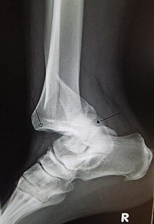
Coxa vara is a deformity of the hip, whereby the angle between the head and the shaft of the femur is reduced to less than 120 degrees. This results in the leg being shortened and the development of a limp. It may be congenital and is commonly caused by injury, such as a fracture. It can also occur when the bone tissue in the neck of the femur is softer than normal, causing it to bend under the weight of the body. This may either be congenital or the result of a bone disorder. The most common cause of coxa vara is either congenital or developmental. Other common causes include metabolic bone diseases, post-Perthes deformity, osteomyelitis, and post traumatic. Shepherd's Crook deformity is a severe form of coxa vara where the proximal femur is severely deformed with a reduction in the neck shaft angle beyond 90 degrees. It is most commonly a sequela of osteogenesis imperfecta, Pagets disease, osteomyelitis, tumour and tumour-like conditions.

Bone fracture Physical damage to the continuity of a bone

A bone fracture is a medical condition in which there is a partial or complete break in the continuity of a bone. In more severe cases, the bone may be broken into several pieces. A bone fracture may be the result of high force impact or stress, or a minimal trauma injury as a result of certain medical conditions that weaken the bones, such as osteoporosis, osteopenia, bone cancer, or osteogenesis imperfecta, where the fracture is then properly termed a pathologic fracture.

Joint dislocation Medical injury

A joint dislocation, also called luxation, occurs when there is an abnormal separation in the joint, where two or more bones meet. A partial dislocation is referred to as a subluxation. Dislocations are often caused by sudden trauma on the joint like an impact or fall. A joint dislocation can cause damage to the surrounding ligaments, tendons, muscles, and nerves. Dislocations can occur in any major joint or minor joint. The most common joint dislocation is a shoulder dislocation.

Arthroplasty is an orthopedic surgical procedure where the articular surface of a musculoskeletal joint is replaced, remodeled, or realigned by osteotomy or some other procedure. It is an elective procedure that is done to relieve pain and restore function to the joint after damage by arthritis or some other type of trauma.

Joint replacement Orthopedic surgery to replace a joint

Replacement arthroplasty, or joint replacement surgery, is a procedure of orthopedic surgery in which an arthritic or dysfunctional joint surface is replaced with an orthopedic prosthesis. Joint replacement is considered as a treatment when severe joint pain or dysfunction is not alleviated by less-invasive therapies. It is a form of arthroplasty, and is often indicated from various joint diseases, including osteoarthritis and rheumatoid arthritis.

Hip dislocation Orthopedic injury

A hip dislocation is when the thighbone (femur) separates from the hip bone (pelvis). Specifically it is when the ball–shaped head of the femur separates from its cup–shaped socket in the hip bone, known as the acetabulum. The joint of the femur and pelvis is very stable, secured by both bony and soft-tissue constraints. With that, dislocation would require significant force which typically results from significant trauma such as from a motor vehicle collision or from a fall from elevation. Hip dislocations can also occur following a hip replacement or from a developmental abnormality known as hip dysplasia.

Madelungs deformity Medical condition

Madelung's deformity is usually characterized by malformed wrists and wrist bones and is often associated with Léri-Weill dyschondrosteosis. It can be bilateral or just in the one wrist. It has only been recognized within the past hundred years. Named after Otto Wilhelm Madelung (1846–1926), a German surgeon, who described it in detail, it was noted by others. Guillaume Dupuytren mentioned it in 1834, Auguste Nélaton in 1847, and Joseph-François Malgaigne in 1855.

Femoral head ostectomy

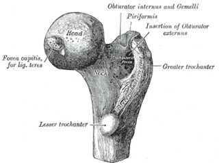
A femoral head ostectomy is a surgical operation to remove the head and neck from the femur. It is performed to alleviate pain, and is a salvage procedure, reserved for condition where pain can not be alleviated in any other way. It is common in veterinary surgery. Other names are excision arthroplasty of the femoral head and neck, Girdlestone's operation, Girdlestone procedure, and femoral head and neck ostectomy.

Ollier disease Medical condition

Ollier disease is a rare sporadic nonhereditary skeletal disorder in which typically benign cartilaginous tumors (enchondromas) develop near the growth plate cartilage. This is caused by cartilage rests that grow and reside within the metaphysis or diaphysis and eventually mineralize over time to form multiple enchondromas. Key signs of the disorder include asymmetry and shortening of the limb as well as an increased thickness of the bone margin. These symptoms are typically first visible during early childhood with the mean age of diagnosis being 13 years of age. Many patients with Ollier disease are prone to develop other malignancies including bone sarcomas that necessitate treatment and the removal of malignant bone neoplasm. Cases in patients with Ollier disease has shown a link to IDH1, IDH2, and PTH1R gene mutations. Currently, there are no forms of treatment for the underlying condition of Ollier disease but complications such as fractures, deformities, malignancies that arise from it can be treated through surgical procedures. The prevalence of this condition is estimated at around 1 in 100,000. It is unclear whether the men or women are more affected by this disorder due to conflicting case studies.

HOSMAT multispecialty Hospital Pvt. Ltd. , the Hospital for Orthopaedics, Sports Medicine, Arthritis & Trauma, is a 350-bed speciality hospital in central Bangalore, India. It also includes Hosmat Joint Replacement Center and HOSMAT Neurosciences. It is currently undergoing expansion to 500 beds, which would make it the largest speciality hospital of its kind in Asia.

Separated shoulder Medical condition

A separated shoulder, also known as acromioclavicular joint injury, is a common injury to the acromioclavicular joint. The AC joint is located at the outer end of the clavicle where it attaches to the acromion of the scapula. Symptoms include non-radiating pain which may make it difficult to move the shoulder. The presence of swelling or bruising and a deformity in the shoulder is also common depending on how severe the dislocation is.

Foot and ankle surgery is a sub-specialty of orthopedics and podiatry that deals with the treatment, diagnosis and prevention of disorders of the foot and ankle. Orthopaedic surgeons are medically qualified, having been through four years of college, followed by 4 years of medical school to obtain an M.D. or D.O. followed by specialist training as a resident in orthopaedics, and only then do they sub-specialise in foot and ankle surgery. Training for a podiatric foot and ankle surgeon consists of four years of college, four years of podiatric medical school (D.P.M.), 3–4 years of a surgical residency and an optional 1 year fellowship.

Protrusio acetabuli is an uncommon defect of the acetabulum, the socket that receives the femoral head to make the hip joint. The hip bone of the pelvic bone/girdle is composed of three bones, the ilium, the ischium and the pubis. In protrusio deformity, there is medial displacement of the femoral head in that the medial aspect of the femoral cortex is medial to the ilioischial line. The socket is too deep and may protrude into the pelvis.

Hip dysplasia Joint abnormality

Hip dysplasia is an abnormality of the hip joint where the socket portion does not fully cover the ball portion, resulting in an increased risk for joint dislocation. Hip dysplasia may occur at birth or develop in early life. Regardless, it does not typically produce symptoms in babies less than a year old. Occasionally one leg may be shorter than the other. The left hip is more often affected than the right. Complications without treatment can include arthritis, limping, and low back pain.

The following outline is provided as an overview of and topical guide to trauma and orthopaedics: