GYPA | |||||||||||||||||||||||||
---|---|---|---|---|---|---|---|---|---|---|---|---|---|---|---|---|---|---|---|---|---|---|---|---|---|
| |||||||||||||||||||||||||
Identifiers | |||||||||||||||||||||||||
Aliases | GYPA , CD235a, GPA, GPErik, GPSAT, HGpMiV, HGpMiXI, HGpSta(C), MN, MNS, PAS-2, glycophorin A (MNS blood group) | ||||||||||||||||||||||||
External IDs | HomoloGene: 48076 GeneCards: GYPA | ||||||||||||||||||||||||
| |||||||||||||||||||||||||
| |||||||||||||||||||||||||
| |||||||||||||||||||||||||
Orthologs | |||||||||||||||||||||||||
Species | Human | Mouse | |||||||||||||||||||||||
Entrez |
| ||||||||||||||||||||||||
Ensembl |
| ||||||||||||||||||||||||
UniProt |
| ||||||||||||||||||||||||
RefSeq (mRNA) |
| ||||||||||||||||||||||||
RefSeq (protein) |
| ||||||||||||||||||||||||
Location (UCSC) | Chr 4: 144.11 – 144.87 Mb | n/a | |||||||||||||||||||||||
PubMed search | [2] | n/a | |||||||||||||||||||||||
Wikidata | |||||||||||||||||||||||||
|
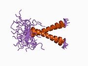
Glycophorin A (MNS blood group), also known as GYPA, is a protein which in humans is encoded by the GYPA gene. [3] GYPA has also recently been designated CD235a (cluster of differentiation 235a).
Proteins are large biomolecules, or macromolecules, consisting of one or more long chains of amino acid residues. Proteins perform a vast array of functions within organisms, including catalysing metabolic reactions, DNA replication, responding to stimuli, providing structure to cells and organisms, and transporting molecules from one location to another. Proteins differ from one another primarily in their sequence of amino acids, which is dictated by the nucleotide sequence of their genes, and which usually results in protein folding into a specific three-dimensional structure that determines its activity.
In biology, a gene is a sequence of nucleotides in DNA or RNA that codes for a molecule that has a function. During gene expression, the DNA is first copied into RNA. The RNA can be directly functional or be the intermediate template for a protein that performs a function. The transmission of genes to an organism's offspring is the basis of the inheritance of phenotypic trait. These genes make up different DNA sequences called genotypes. Genotypes along with environmental and developmental factors determine what the phenotypes will be. Most biological traits are under the influence of polygenes as well as gene–environment interactions. Some genetic traits are instantly visible, such as eye color or number of limbs, and some are not, such as blood type, risk for specific diseases, or the thousands of basic biochemical processes that constitute life.
The cluster of differentiation is a protocol used for the identification and investigation of cell surface molecules providing targets for immunophenotyping of cells. In terms of physiology, CD molecules can act in numerous ways, often acting as receptors or ligands important to the cell. A signal cascade is usually initiated, altering the behavior of the cell. Some CD proteins do not play a role in cell signaling, but have other functions, such as cell adhesion. CD for humans is numbered up to 371.
Glycophorins A (GYPA; this protein) and B (GYPB) are major sialoglycoproteins of the human erythrocyte membrane which bear the antigenic determinants for the MN and Ss blood groups. In addition to the M or N and S or s antigens, that commonly occur in all populations, about 40 related variant phenotypes have been identified. These variants include all the variants of the Miltenberger complex and several isoforms of Sta; also, Dantu, Sat, He, Mg, and deletion variants Ena, S-s-U- and Mk. Most of the variants are the result of gene recombinations between GYPA and GYPB. [3]
Glycophorin B also known as sialoglycoprotein delta and SS-active sialoglycoprotein is a protein which in humans is encoded by the GYPB gene. GYPB has also recently been designated CD235b.
GypA, GypB and GypE are members of the same family and are located on the long arm of chromosome 4 (chromosome 4q31). The family evolved via two separate gene duplication events. The initial duplication gave rise to two genes one of subsequently evolved into GypA and the other which give rise via a second duplication event to GypB and GypE. These events appear to have occurred within a relatively short time span. The second duplication appears to have occurred via an unequal crossing over event.
The GypA gene itself consists of 7 exons and has 97% sequence homology with GypB and GypE from the 5' untranslated transcription region (UTR) to the coding sequence encoding the first 45 amino acids. The exon at this point encodes the transmembrane domain. Within the intron downstream of this pint is an Alu repeat. The cross over event which created the genes ancestral to GypA and GypB/E occurred within this region.
GypA can be found in all primates. GypB can be found only in gorillas and some of the higher primates suggesting that the duplication events occurred only recently.
Gorillas are ground-dwelling, predominantly herbivorous apes that inhabit the forests of central Sub-Saharan Africa. The genus Gorilla is divided into two species: the eastern gorillas and the western gorillas, and either four or five subspecies. They are the largest living primates. The DNA of gorillas is highly similar to that of humans, from 95 to 99% depending on what is included, and they are the next closest living relatives to humans after the chimpanzees and bonobos.
There are about one million copies of this protein per erythrocyte. [Reference needed]
The MNS blood group was the second set of antigens discovered. M and N were identified in 1927 by Landsteiner and Levine. S and s in were described later in 1947.
The frequencies of these antigens are
White people is a racial classification specifier, used mostly and often exclusively for people of European descent; depending on context, nationality, and point of view. The term has at times been expanded to encompass persons of Middle Eastern and North African descent, persons who are often considered non-white in other contexts. The usage of "white people" or a "white race" as nomenclature for European populations - indicated by their light skin, among other physical characteristics, and contrasting with "black people", Amerindians, and other "colored" people or "persons of color" - originated in the 17th century. It was only during the 19th century that the category of white people was transformed in a quasi-scientific system of race and skin color relations. The term "Caucasian" is sometimes used as a synonym for "white" in its racial sense and sometimes to refer to a larger racial category that includes white people among other groups.
Negroid is a grouping of human beings historically regarded as a biological taxon. The term has been used by forensic and physical anthropologists to refer to individuals and populations that share certain morphological and skeletal traits that are frequent among populations in most of Sub-Saharan Africa and isolated parts of South and Southeast Asia (Negritos). Within Africa, a racial dividing line separating Caucasoid physical types from Negroid physical types was held to have existed, with Negroid groups forming most of the population south of the area which stretched from the southern Sahara desert in the west to the African Great Lakes in the southeast.
The M and N antigens differ at two amino acid residues: the M allele has serine at position 1 (C at nucleotide 2) and glycine at position 5 (G at nucleotide 14) while the N allele has leucine at position 1 (T at nucleotide 2) and glutamate at position 5 (A at nucleotide 14). Both glycophorin A and B bind the Vicia graminea anti-N lectin.
There are about 40 known variants in the MNS blood group system. These have arisen largely as a result of mutations within the 4 kb region coding for the extracellular domain. These include the antigens Mg, Dantu, Henshaw (He), Miltenberger, Nya, Osa, Orriss (Or), Raddon (FR) and Stones (Sta). Chimpanzees also have an MN blood antigen system. [4] In chimpanzees M reacts strong but N only weakly.
In individuals who lack both glycophorin A and B the phenotype has been designated Mk. [5]
The Dantu antigen was described in 1984. [6] The Dantu antigen has an apparent molecular weight of 29 kiloDaltons (kDa) and 99 amino acids. The first 39 amino acids of the Dantu antigen are derived from glycophorin B and residues 40-99 are derived from glycophorin A. Dantu is associated with very weak s antigen, a protease-resistant N antigen and either very weak or no U antigen. There are at least three variants: MD, NE and Ph. [7] The Dantu phenotype occurs with a frequency of Dantu phenotype is ~0.005 in American Blacks and < 0.001 in Germans. [8]
The Henshaw (He) antigen is due to a mutation of the N terminal region. There are three differences in the first three amino acid residues: the usual form has Tryptophan 1-Serine-Threonine-Serine-Glycine 5 while Henshaw has Leucine 1-Serine-Threonine-Threonine-Glutamate 5. This antigen is rare in Caucasians but occurs at a frequency of 2.1% in US and UK of African origin. It occurs at the rate of 7.0% in blacks in Natal [9] and 2.7% in West Africans. [10] At least 3 variants of this antigen have been identified.
The Miltenberger (Mi) subsystem originally consisting of five phenotypes (Mia, Vw, Mur, Hil and Hut) [11] now has 11 recognised phenotypes numbered I to XI (The antigen 'Mur' is named after to the patient the original serum was isolated from - a Mrs Murrel.) The name originally given to this complex refers to the reaction erythrocytes gave to the standard Miltenberger antisera used to test them. The subclasses were based on additional reactions with other standard antisera.
Mi-I (Mia), Mi-II(Vw), Mi-VII and Mi-VIII are carried on glycophorin A. Mi-I is due to a mutation at amino acid 28 (threonine to methionine: C→T at nucleotide 83) resulting in a loss of the glycosylation at the asparagine26 residue. [12] [13] Mi-II is due to a mutation at amino acid 28 (threonine to lysine:C->A at nucleotide 83). [13] Similar to the case of Mi-I this mutation results in a loss of the glycosylation at the asparagine 26 residue. This alteration in glycoslation is detectable by the presence of a new 32kDa glycoprotein stainable with PAS. [14] Mi-VII is due to a double mutation in glycophorin A converting an arginine residue into a threonine residue and a tyrosine residue into a serine at the positions 49 and 52 respectively. [15] The threonine-49 residue is glycosylated. This appears to be the origin of one of the Mi-VII specific antigens (Anek) which is known to lie between residues 40-61 of glycophorin A and comprises sialic acid residue(s) attached to O-glycosidically linked oligosaccharide(s). This also explains the loss of a high frequency antigen ((EnaKT)) found in normal glycophorin A which is located within the residues 46-56. Mi-VIII is due to a mutation at amino acid residue 49 (arginine->threonine). [16] M-VIII shares the Anek determinant with MiVII. [17] Mi-III, Mi-VI and Mi-X are due to rearrangements of glycophorin A and B in the order GlyA (alpha)-GlyB (delta)-GlyA (alpha). [18] Mil-IX in contrast is a reverse alpha-delta-alpha hybrid gene. [19] Mi-V, MiV(J.L.) and Sta are due to unequal but homologous crossing-over between alpha and delta glycophorin genes. [20] The MiV and MiV(J.L.) genes are arranged in the same 5' alpha-delta 3' frame whereas Sta gene is in a reciprocal 5'delta-alpha 3' configuration.
The incidence of Mi-I in Thailand is 9.7%. [21]
Peptide constructs representative of Mia mutations MUT and MUR have been attached onto red blood cells (known as kodecytes) and are able to detect antibodies against these Miltenberger antigens [22] [23] [24]
Although uncommon in Caucasians (0.0098%) and Japanese (0.006%), the frequency of Mi-III is exceptionally high in several Taiwanese aboriginal tribes (up to 90%). In contrast its frequency is 2-3% in Han Taiwanese (Minnan). The Mi-III phenotype occurs in 6.28% of Hong Kong Chinese. [25]
Mi-IX (MNS32) occurs with a frequency of 0.43% in Denmark. [26]
Stones (Sta) has been shown to be the product of a hybrid gene of which the 5'-half is derived from the glycophorin B whereas the 3'-half is derived from the glycophorin A. Several isoforms are known. This antigen is now considered to be part of the Miltenberger complex.
A related antigen is Sat. This gene has six exons of which exon I to exon IV are identical to the N allele of glycophorin A whereas its 3' portion, including exon V and exon VI, are derived from the glycophorin B gene. The mature protein SAT protein contains 104 amino acid residues.
Orriss (Or) appears to be a mutant of glyphorin A but its precise nature has not yet been determined. [27]
The Mg antigen is carried on glycophorin A and lacks three O-glycolated side chains. [28]
Osa (MNS38) is due to a mutation at nucleotide 273 (C->T) lying within exon 3 resulting in the replacement of a proline residue with a serine. [29]
Nya (MNS18) is due to a mutation at nucleotide 194 (T->A) which results in the substitution of an aspartate residue with a glutamate. [29]
Anti-M although occurring naturally has rarely been implicated in transfusion reactions. Anti-N is not considered to cause transfusion reactions. Severe reactions have been reported with anti-Miltenberger. Anti Mi-I (Vw) and Mi-III has been recognised as a cause of haemolytic disease of the newborn. [30] Raddon has been associated with severe transfusion reactions. [31]
The Wright b antigen (Wrb) is located on glycophorin A and acts as a receptor for the malaria parasite Plasmodium falciparum . [32] Cells lacking glycophorins A (Ena) are resistant to invasion by this parasite. [33]
The erythrocyte binding antigen 175 of P. falciparum recognises the terminal Neu5Ac(alpha 2-3)Gal-sequences of glycophorin A. [34]
Several viruses bind to glycophorin A including hepatitis A virus (via its capsid), [35] bovine parvovirus , [36] Sendai virus , [37] influenza A and B , [38] group C rotavirus , [39] encephalomyocarditis virus [40] and reovirus es. [41]
Glycophorin C plays a functionally important role in maintaining erythrocyte shape and regulating membrane material properties, possibly through its interaction with protein 4.1. Moreover, it has previously been shown that membranes deficient in protein 4.1 exhibit decreased content of glycophorin C. It is also an integral membrane protein of the erythrocyte and acts as the receptor for the Plasmodium falciparum protein PfEBP-2.
Duffy antigen/chemokine receptor (DARC), also known as Fy glycoprotein (FY) or CD234, is a protein that in humans is encoded by the DARC gene.
Complement receptor type 1 (CR1) also known as C3b/C4b receptor or CD35 is a protein that in humans is encoded by the CR1 gene.
CD36, also known as platelet glycoprotein 4, fatty acid translocase (FAT), scavenger receptor class B member 3 (SCARB3), and glycoproteins 88 (GP88), IIIb (GPIIIB), or IV (GPIV) is a protein that in humans is encoded by the CD36 gene. The CD36 antigen is an integral membrane protein found on the surface of many cell types in vertebrate animals. It imports fatty acids inside cells and is a member of the class B scavenger receptor family of cell surface proteins. CD36 binds many ligands including collagen, thrombospondin, erythrocytes parasitized with Plasmodium falciparum, oxidized low density lipoprotein, native lipoproteins, oxidized phospholipids, and long-chain fatty acids.
The ABO blood group system is used to denote the presence of one, both, or neither of the A and B antigens on erythrocytes. In human blood transfusions it is the most important of the 36 different blood type classification systems currently recognized. A very rare mismatch in this, or any other serotype, can cause a serious, potentially fatal, adverse reaction after a transfusion, or a contra-indicated immune response to an organ transplant. The associated anti-A and anti-B antibodies are usually IgM antibodies, which are produced in the first years of life by sensitization to environmental substances, such as food, bacteria, and viruses.
The Kell antigen system is a group of antigens on the human red blood cell surface which are important determinants of blood type and are targets for autoimmune or alloimmune diseases which destroy red blood cells. Kell can be noted as K, k, or Kp. The Kell antigens are peptides found within the Kell protein, a 93-kilodalton transmembrane zinc-dependent endopeptidase which is responsible for cleaving endothelin-3.
McLeod syndrome is an X-linked recessive genetic disorder that may affect the blood, brain, peripheral nerves, muscle, and heart. It is caused by a variety of recessively inherited mutations in the XK gene on the X chromosome. The gene is responsible for producing the Kx protein, a secondary supportive protein for the Kell antigen on the red blood cell surface.
Animal erythrocytes have cell surface antigens that undergo polymorphism and give rise to blood types. Antigens from the human ABO blood group system are also found in apes and Old World monkeys, and the system traces back to the origin of hominoids. Other animal blood sometimes agglutinates with human blood group reagents, but the structure of the blood group antigens in animals is not always identical to those typically found in humans. The classification of most animal blood groups therefore uses different blood typing systems to those used for classification of human blood.
The MNS antigen system is a human blood group system based upon two genes on chromosome 4. There are currently 46 antigens in the system, but the five most important are called M, N, S, s, and U.
The LW blood system was first described by Landsteiner and Wiener in 1940. It was often confused with the Rh system, not becoming a separate antigen system until 1982. The LW and RhD antigens are genetically independent though they are phenotypically related and the LW antigen is expressed more strongly on RhD positive cells than on RhD negative cells. In most populations, the antithetical LW antigens, LWa and LWb are present as very high and very low frequency, respectively.
Galactoside 3(4)-L-fucosyltransferase is an enzyme that in humans is encoded by the FUT3 gene.
Blood group Rh(CE) polypeptide is a protein that in humans is encoded by the RHCE gene. RHCE has also recently been designated CD240CE.
Rh-associated glycoprotein (RHAG) is an ammonia transporter protein that in humans is encoded by the RHAG gene. RHAG has also recently been designated CD241. Mutations in the RHAG gene can cause stomatocytosis.
Ecto-ADP-ribosyltransferase 4 is an enzyme that in humans is encoded by the ART4 gene. ART4 has also been designated as CD297.
Erythroid membrane-associated protein is a protein that in humans is responsible for the Scianna blood group system, and is encoded by the ERMAP gene.
Glycophorin-E is a protein that in humans is encoded by the GYPE gene.
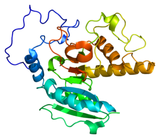
Histo-blood group ABO system transferase is an enzyme with glycosyltransferase activity, which is encoded by the ABO gene in humans. It is ubiquitously expressed in many tissues and cell types. ABO determines the ABO blood group of an individual by modifying the oligosaccharides on cell surface glycoproteins. Variations in the sequence of the protein between individuals determine the type of modification and the blood group. The ABO gene also contains one of 27 SNPs associated with increased risk of coronary artery disease.
The BGMUT Database documents allelic variations in the genes encoding for human blood group systems. It was set up in 1999 through an initiative of the Human Genome Variation Society (HGVS). Since 2006, it has been a part of the dbRBC resource of NCBI at the NIH. In addition to being a repository of the genetic variations of the blood group antigen-encoding genes, the database also provides information on the blood group systems, the genes that encode them, the serological phenotypes associated with the alleles of the genes, etc. Information on genetic variations in some non-human orthologous genes is also provided.
Rh blood group, D antigen also known as Rh polypeptide 1 (RhPI) or cluster of differentiation 240D (CD240D) is a protein that in humans is encoded by the RHD gene.
This article incorporates text from the United States National Library of Medicine, which is in the public domain.