ACTG1 | |||||||||||||||||||||||||||||||||||||||||||||||||||
---|---|---|---|---|---|---|---|---|---|---|---|---|---|---|---|---|---|---|---|---|---|---|---|---|---|---|---|---|---|---|---|---|---|---|---|---|---|---|---|---|---|---|---|---|---|---|---|---|---|---|---|
| |||||||||||||||||||||||||||||||||||||||||||||||||||
Identifiers | |||||||||||||||||||||||||||||||||||||||||||||||||||
Aliases | ACTG1 , ACT, ACTG, BRWS2, DFNA20, DFNA26, HEL-176, actin gamma 1 | ||||||||||||||||||||||||||||||||||||||||||||||||||
External IDs | OMIM: 102560; MGI: 87906; HomoloGene: 74402; GeneCards: ACTG1; OMA:ACTG1 - orthologs | ||||||||||||||||||||||||||||||||||||||||||||||||||
| |||||||||||||||||||||||||||||||||||||||||||||||||||
| |||||||||||||||||||||||||||||||||||||||||||||||||||
| |||||||||||||||||||||||||||||||||||||||||||||||||||
| |||||||||||||||||||||||||||||||||||||||||||||||||||
| |||||||||||||||||||||||||||||||||||||||||||||||||||
Wikidata | |||||||||||||||||||||||||||||||||||||||||||||||||||
|
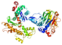
Actin, cytoplasmic 2, or gamma-actin is a protein that in humans is encoded by the ACTG1 gene. [5] Gamma-actin is widely expressed in cellular cytoskeletons of many tissues; in adult striated muscle cells, gamma-actin is localized to Z-discs and costamere structures, which are responsible for force transduction and transmission in muscle cells. Mutations in ACTG1 have been associated with nonsyndromic hearing loss and Baraitser-Winter syndrome, as well as susceptibility of adolescent patients to vincristine toxicity.
Human gamma-actin is 41.8 kDa in molecular weight and 375 amino acids in length. [6] Actins are highly conserved proteins that are involved in various types of cell motility, and maintenance of the cytoskeleton. In vertebrates, three main groups of actin paralogs, alpha, beta, and gamma, have been identified. [7]
The alpha actins are found in muscle tissues and are a major constituent of the sarcomere contractile apparatus. The beta and gamma actins co-exist in most cell types as components of the cytoskeleton, and as mediators of internal cell motility. Actin, gamma 1, encoded by this gene, is found in non-muscle cells in the cytoplasm, and in muscle cells at costamere structures, or transverse points of cell-cell adhesion that run perpendicular to the long axis of myocytes. [8] [9] [10]
In myocytes, sarcomeres adhere to the sarcolemma via costameres, which align at Z-discs and M-lines. [11] The two primary cytoskeletal components of costameres are desmin intermediate filaments and gamma-actin microfilaments. [12] It has been shown that gamma-actin interacting with another costameric protein dystrophin is critical for costameres forming mechanically strong links between the cytoskeleton and the sarcolemmal membrane. [13] [14] Additional studies have shown that gamma-actin colocalizes with alpha-actinin and GFP-labeled gamma actin localized to Z-discs, whereas GFP-alpha-actin localized to pointed ends of thin filaments, indicating that gamma actin specifically localizes to Z-discs in striated muscle cells. [15] [16] [17]
During development of myocytes, gamma actin is thought to play a role in the organization and assembly of developing sarcomeres, evidenced in part by its early colocalization with alpha-actinin. [18] Gamma-actin is eventually replaced by sarcomeric alpha-actin isoforms, [19] [20] [21] with low levels of gamma-actin persisting in adult myocytes which associate with Z-disc and costamere domains. [15] [22] [23]
Insights into the function of gamma-actin in muscle have come from studies employing transgenesis. In a skeletal muscle-specific knockout of gamma-actin in mice, these animals showed no detectable abnormalities in development; however, knockout mice showed muscle weakness and fiber necrosis, along with decreased isometric twitch force, disrupted intrafibrillar and interfibrillar connections among myocytes, and myopathy. [24]
An autosomal dominant mutation in ACTG1 in the DFNA20/26 locus at 17q25-qter was identified in patients with hearing loss. A Thr278Ile mutation was identified in helix 9 of gamma-actin protein, which is predicted to alter protein structure. This study identified the first disease causing mutation in gamma-actin and underlies the importance of gamma-actin as structural elements of the inner ear hair cells. [25] Since then, other ACTG1 mutations have been linked to nonsyndromic hearing loss, including Met305Thr. [26]
A missense mutation in ACTG1 at Ser155Phe has also been identified in patients with Baraitser-Winter syndrome, which is a developmental disorder characterized by congenital ptosis, excessively-arched eyebrows, hypertelorism, ocular colobomata, lissencephaly, short stature, seizures and hearing loss. [27] [28]
Differential expression of ACTG1 mRNA was also identified in patients with Sporadic Amyotrophic Lateral Sclerosis, a devastating disease with unknown causality, using a sophisticated bioinformatics approach employing Affymetrix long-oligonucleotide BaFL methods. [29]
Single nucleotide polymorphisms in ACTG1 have been associated with vincristine toxicity, which is part of the standard treatment regimen for childhood acute lymphoblastic leukemia. Neurotoxicity was more frequent in patients that were ACTG1 Gly310Ala mutation carriers, suggesting that this may play a role in patient outcomes from vincristine treatment. [30]
ACTG1 has been shown to interact with:
The cytoskeleton is a complex, dynamic network of interlinking protein filaments present in the cytoplasm of all cells, including those of bacteria and archaea. In eukaryotes, it extends from the cell nucleus to the cell membrane and is composed of similar proteins in the various organisms. It is composed of three main components: microfilaments, intermediate filaments, and microtubules, and these are all capable of rapid growth or disassembly depending on the cell's requirements.
Actin is a family of globular multi-functional proteins that form microfilaments in the cytoskeleton, and the thin filaments in muscle fibrils. It is found in essentially all eukaryotic cells, where it may be present at a concentration of over 100 μM; its mass is roughly 42 kDa, with a diameter of 4 to 7 nm.
Intermediate filaments (IFs) are cytoskeletal structural components found in the cells of vertebrates, and many invertebrates. Homologues of the IF protein have been noted in an invertebrate, the cephalochordate Branchiostoma.
In mammalian cells, vinculin is a membrane-cytoskeletal protein in focal adhesion plaques that is involved in linkage of integrin adhesion molecules to the actin cytoskeleton. Vinculin is a cytoskeletal protein associated with cell-cell and cell-matrix junctions, where it is thought to function as one of several interacting proteins involved in anchoring F-actin to the membrane.
Spectrin is a cytoskeletal protein that lines the intracellular side of the plasma membrane in eukaryotic cells. Spectrin forms pentagonal or hexagonal arrangements, forming a scaffold and playing an important role in maintenance of plasma membrane integrity and cytoskeletal structure. The hexagonal arrangements are formed by tetramers of spectrin subunits associating with short actin filaments at either end of the tetramer. These short actin filaments act as junctional complexes allowing the formation of the hexagonal mesh. The protein is named spectrin since it was first isolated as a major protein component of human red blood cells which had been treated with mild detergents; the detergents lysed the cells and the hemoglobin and other cytoplasmic components were washed out. In the light microscope the basic shape of the red blood cell could still be seen as the spectrin-containing submembranous cytoskeleton preserved the shape of the cell in outline. This became known as a red blood cell "ghost" (spectre), and so the major protein of the ghost was named spectrin.
Plectin is a giant protein found in nearly all mammalian cells which acts as a link between the three main components of the cytoskeleton: actin microfilaments, microtubules and intermediate filaments. In addition, plectin links the cytoskeleton to junctions found in the plasma membrane that structurally connect different cells. By holding these different networks together, plectin plays an important role in maintaining the mechanical integrity and viscoelastic properties of tissues.
Paxillin is a protein that in humans is encoded by the PXN gene. Paxillin is expressed at focal adhesions of non-striated cells and at costameres of striated muscle cells, and it functions to adhere cells to the extracellular matrix. Mutations in PXN as well as abnormal expression of paxillin protein has been implicated in the progression of various cancers.
The costamere is a structural-functional component of striated muscle cells which connects the sarcomere of the muscle to the cell membrane.
Protein 4.1,, is a protein associated with the cytoskeleton that in humans is encoded by the EPB41 gene. Protein 4.1 is a major structural element of the erythrocyte membrane skeleton. It plays a key role in regulating membrane physical properties of mechanical stability and deformability by stabilizing spectrin-actin interaction. Protein 4.1 interacts with spectrin and short actin filaments to form the erythrocyte membrane skeleton. Mutations of spectrin and protein 4.1 are associated with elliptocytosis or spherocytosis and anemia of varying severity.
Integrin beta-1 (ITGB1), also known as CD29, is a cell surface receptor that in humans is encoded by the ITGB1 gene. This integrin associates with integrin alpha 1 and integrin alpha 2 to form integrin complexes which function as collagen receptors. It also forms dimers with integrin alpha 3 to form integrin receptors for netrin 1 and reelin. These and other integrin beta 1 complexes have been historically known as very late activation (VLA) antigens.
Dystrobrevin is a protein that binds to dystrophin in the costamere of skeletal muscle cells. In humans, there are at least two isoforms of dystrobrevin, dystrobrevin alpha and dystrobrevin beta.
Actin, alpha skeletal muscle is a protein that in humans is encoded by the ACTA1 gene.
Actin beta is one of six different actin isoforms which have been identified in humans. This is one of the two nonmuscle cytoskeletal actins. Actins are highly conserved proteins that are involved in cell motility, structure and integrity. Alpha actins are a major constituent of the contractile apparatus.
Alpha-actinin-2 is a protein which in humans is encoded by the ACTN2 gene. This gene encodes an alpha-actinin isoform that is expressed in both skeletal and cardiac muscles and functions to anchor myofibrillar actin thin filaments and titin to Z-discs.
Filamin-C (FLN-C) also known as actin-binding-like protein (ABPL) or filamin-2 (FLN2) is a protein that in humans is encoded by the FLNC gene. Filamin-C is mainly expressed in cardiac and skeletal muscles, and functions at Z-discs and in subsarcolemmal regions.
Actin, gamma-enteric smooth muscle is a protein that in humans is encoded by the ACTG2 gene.
Myotilin is a protein that in humans is encoded by the MYOT gene. Myotilin also known as TTID is a muscle protein that is found within the Z-disc of sarcomeres.
Cysteine and glycine-rich protein 3 also known as cardiac LIM protein (CLP) or muscle LIM protein (MLP) is a protein that in humans is encoded by the CSRP3 gene.
Talin-1 is a protein that in humans is encoded by the TLN1 gene. Talin-1 is ubiquitously expressed, and is localized to costamere structures in cardiac and skeletal muscle cells, and to focal adhesions in smooth muscle and non-muscle cells. Talin-1 functions to mediate cell-cell adhesion via the linkage of integrins to the actin cytoskeleton and in the activation of integrins. Altered expression of talin-1 has been observed in patients with heart failure, however no mutations in TLN1 have been linked with specific diseases.
Talin-2 is a protein in humans that is encoded by the TLN2 gene. It belongs to the talin protein family. This gene encodes a protein related to talin-1, a cytoskeletal protein that plays a significant role in the assembly of actin filaments. Talin-2 is expressed at high levels in cardiac muscle and functions to provide linkages between the extracellular matrix and actin cytoskeleton at costamere structures to transduce force laterally.