Surfer's ear | |
---|---|
Exostoses in the ear canal, as seen through otoscopy | |
Specialty | ENT surgery |
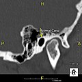
Surfer's ear is the common name for an exostosis or abnormal bone growth within the ear canal. They are otherwise benign hyperplasias (growths) of the tympanic bone thought to be caused by frequent cold-water exposure. [1] Cases are often asymptomatic. [1] Surfer's ear is not the same as swimmer's ear, although infection can result as a side effect.
Irritation from cold wind and water exposure causes the bone surrounding the ear canal to develop lumps of new bony growth which constrict the ear canal. Where the ear canal is actually blocked by this condition, water and wax can become trapped and give rise to infection. The condition is so named due to its high prevalence among cold water surfers, although it can occur in any water temperature due to the evaporative cooling caused by wind and the presence of water in the ear canal.
Most avid surfers have at least some mild bone growths, causing little to no problems. [2] The condition is gradually progressive and can generally be prevented by shielding the ear from water by consistently using earplugs and wetsuit hoods. The condition is not limited to surfing and can occur in any activity with cold, wet, windy conditions such as windsurfing, kayaking, sailing, jet skiing, kitesurfing, and diving.
In general, one ear will be somewhat worse than the other due to the prevailing wind direction of the area surfed [3] or the side that most often strikes the wave first. Common symptoms include recurrent otitis externa, obstructed sensation, conductive hearing loss, pain in the ear, and tinnitus. [1]
The majority of patients present in their mid-30s to late 40s. This is likely due to a combination of the slow growth of the bone and the decreased participation in activities associated with surfer's ear past the 30s. However, surfer's ear is possible at any age and is directly proportional to the amount of time spent in cold, wet, windy weather without adequate protection. [4]
The normal ear canal is approximately 7 mm in diameter and has a volume of approximately 0.8 ml (approximately one-sixth of a teaspoon). [5] As the condition progresses, the diameter narrows and can even close completely if untreated, although people generally seek help once the passage has constricted to 0.5–2 mm due to the noticeable hearing impairment. While not necessarily harmful in and of itself, constriction of the ear canal from these growths can trap debris, leading to painful and difficult to treat infections.
A 2015 review of eight other studies found the prevalence ranged from 31% to 80%, largely depending on sea temperature of the geographical area studied, with colder climates having higher prevalence. [6] [7] The study also found among 105 surfers in Cornwall, England 63.8% had surfer's ear. [6]
The widespread use of wetsuits has allowed people to surf in much colder waters, which has increased the incidence and severity of surfer's ear for people who do not properly protect their ears. [4] Preventative measures include avoiding activity during extremely cold or windy conditions, and keep the ear canal as warm and dry as possible through the use of earplugs and wetsuit hoods. [8] In one study, two thirds of 92 surfers were aware that surfer's ear could be prevented. [8] One study suggests that for kayakers, since surfer's ear is a slowly progressive condition, kayakers may not wear earplugs until syptoms appear. [1]
Outside of symptom relief, the only treatment is surgical removal of the formed exostoses. [9] Although not an especially hazardous surgery, exostosis removal can result in major complications such as facial nerve paralysis, canal stenosis, temporomandibular joint prolapse, hearing loss, persistent deep bony lip, and persistent eardrum perforations. [9]
Surgical techniques include drilling the bone off and later cleaning debris using a osteotome, typically under local anesthesia or general anesthesia with the use of a microscope. [10] [11] [12] After surgery it is recommended to continue keeping the ear clear of water, and for one study using ciprofloxacin/hydrocortisone drops were recommended for one week post-surgery to avoid infection, and 90% of the study group had fully healed after four weeks. [10]
Archeological research in Gran Canaria, Spain, has found a relatively high prevalence of exostosis among Pre-Hispanic craniums, reaching 34.35% in coastal burial places. Not all coastal craniums presented exostosis but there were no differences between sexes. Researchers thus proposed a social division of work among the Canarii, with certain individuals, male or female, specializing in fishing by immersion and swimming. [13]
Cholesteatoma is a destructive and expanding growth consisting of keratinizing squamous epithelium in the middle ear and/or mastoid process. Cholesteatomas are not cancerous as the name may suggest, but can cause significant problems because of their erosive and expansile properties. This can result in the destruction of the bones of the middle ear (ossicles), as well as growth through the base of the skull into the brain. They often become infected and can result in chronically draining ears. Treatment almost always consists of surgical removal.
The Eustachian tube, also called the auditory tube or pharyngotympanic tube, is a tube that links the nasopharynx to the middle ear, of which it is also a part. In adult humans, the Eustachian tube is approximately 35 mm (1.4 in) long and 3 mm (0.12 in) in diameter. It is named after the sixteenth-century Italian anatomist Bartolomeo Eustachi.
Earwax, also known by the medical term cerumen, is a waxy substance secreted in the ear canal of humans and other mammals. Earwax can be many colors, including brown, orange, red, yellowish, and gray. Earwax protects the skin of the human ear canal, assists in cleaning and lubrication, and provides protection against bacteria, fungi, particulate matter, and water.
Stapedectomy is a surgical procedure in which the stapes bone is removed from the middle ear and replaced with a prosthesis.
A myringotomy is a surgical procedure in which an incision is created in the eardrum to relieve pressure caused by excessive buildup of fluid, or to drain pus from the middle ear. A tympanostomy tube may be inserted through the eardrum to keep the middle ear aerated for a prolonged time and to prevent reaccumulation of fluid. Without the insertion of a tube, the incision usually heals spontaneously within two to three weeks. Depending on the type, the tube is either naturally extruded in 6 to 12 months or removed during a minor procedure.
Unilateral hearing loss (UHL) is a type of hearing impairment where there is normal hearing in one ear and impaired hearing in the other ear.
An exostosis, also known as a bone spur, is the formation of new bone on the surface of a bone. Exostoses can cause chronic pain ranging from mild to debilitatingly severe, depending on the shape, size, and location of the lesion. It is most commonly found in places like the ribs, where small bone growths form, but sometimes larger growths can grow on places like the ankles, knees, shoulders, elbows and hips. Very rarely are they on the skull.
A bone-anchored hearing aid (BAHA) is a type of hearing aid based on bone conduction. It is primarily suited for people who have conductive hearing losses, unilateral hearing loss, single-sided deafness and people with mixed hearing losses who cannot otherwise wear 'in the ear' or 'behind the ear' hearing aids. They are more expensive than conventional hearing aids, and their placement involves invasive surgery which carries a risk of complications, although when complications do occur, they are usually minor.
A buccal exostosis is an exostosis on the buccal surface of the alveolar ridge of the maxilla or mandible. More commonly seen in the maxilla than the mandible, buccal exostoses are considered to be site specific. Existing as asymptomatic bony nodules, buccal exostoses don’t usually present until adult life, and some consider buccal exostoses to be a variation of normal anatomy rather than disease. Bone is thought to become hyperplastic, consisting of mature cortical and trabecular bone with a smooth outer surface. They are less common when compared with mandibular tori.
Geniculate ganglionitis or geniculate neuralgia (GN), also called nervus intermedius neuralgia, Ramsay Hunt syndrome, or Hunt's neuralgia, is a rare disorder characterized by severe paroxysmal neuralgic pain deep in the ear, that may spread to the ear canal, outer ear, mastoid or eye regions. GN may also occur in combination with trigeminal or glossopharyngeal neuralgia.
Tympanosclerosis is a condition caused by hyalinization and subsequent calcification of subepithelial connective tissue of the tympanic membrane and middle ear, sometimes resulting in a detrimental effect to hearing.
Otitis externa, also called swimmer's ear, is inflammation of the ear canal. It often presents with ear pain, swelling of the ear canal, and occasionally decreased hearing. Typically there is pain with movement of the outer ear. A high fever is typically not present except in severe cases.
Ceruminous adenocarcinoma is a malignant neoplasm derived from ceruminous glands of the external auditory canal. This tumor is rare, with several names used in the past. Synonyms have included cylindroma, ceruminoma, ceruminous adenocarcinoma, not otherwise specified (NOS), ceruminous adenoid cystic carcinoma (ACC), and ceruminous mucoepidermoid carcinoma.
An otic polyp is a benign proliferation of chronic inflammatory cells associated with granulation tissue, in response to a longstanding inflammatory process of the middle ear.
Endoscopic ear surgery (EES) is a minimally invasive alternative to traditional ear surgery and is defined as the use of the rigid endoscope, as opposed to a surgical microscope, to visualize the middle and inner ear during otologic surgery. During endoscopic ear surgery the surgeon holds the endoscope in one hand while working in the ear with the other. To allow this kind of single-handed surgery, different surgical instruments have to be used. Endoscopic visualization has improved due to high-definition video imaging and wide-field endoscopy, and being less invasive, EES is gaining importance as an adjunct to microscopic ear surgery.
Cartilage conduction is a pathway by which sound signals are transmitted to the inner ear. In 2004, Hiroshi Hosoi discovered this pathway and named “cartilage conduction”. Hearing by cartilage conduction is distinct from conventional sound-conduction pathways, such as air or bone, because it is realized by touching a transducer on the aural cartilage and does not involve the vibration of the skull bone. Therefore, cartilage conduction is referred to as the “third auditory pathway”.
Granular myringitis is a long term condition in which there is inflammation of the tympanic membrane in the ear and formation of granulation tissue within the tympanic membrane. It is a type of otitis externa.
Eustachian tube dysfunction (ETD) is a disorder where pressure abnormalities in the middle ear result in symptoms.
Keratosis obturans is a relatively uncommon ear disease, where a dense plug of keratin, formed by abnormal accumulation of desquamated skin in sheet like layers (lamellae), forms in the bony (deeper) part of the external auditory canal. It is clinically diagnosed when removal of the debris shows silvery white peripheral matrix and causes excruciating pain.
LaVonne Bernadene Bergstrom was an American surgeon, otolaryngologist, and medical school professor. She was president of the American Auditory Society in 1987.