Filaggrin

Last updated
FLG
FLG protein.png
Available structures
PDB Human UniProt search: PDBe RCSB
Identifiers
Aliases FLG , ATOD2, filaggrin
External IDs OMIM: 135940; HomoloGene: 136751; GeneCards: FLG; OMA:FLG - orthologs
Orthologs
SpeciesHumanMouse
Entrez
Ensembl
UniProt
RefSeq (mRNA)

NM_002016

n/a

RefSeq (protein)

NP_002007

n/a

Location (UCSC) Chr 1: 152.3 – 152.33 Mb n/a
PubMed search [2] n/a
Wikidata
View/Edit Human

Filaggrin (filament aggregating protein) is a filament-associated protein that binds to keratin fibers in epithelial cells. Ten to twelve filaggrin units are post-translationally hydrolyzed from a large profilaggrin precursor protein during terminal differentiation of epidermal cells. [3] In humans, profilaggrin is encoded by the FLG gene, which is part of the S100 fused-type protein (SFTP) family within the epidermal differentiation complex on chromosome 1q21. [4] In cetaceans and sirenians, the FLG family has lost its function, with the curious exception of manatees in the latter clade: manatees still retain some functional FLG genes. [5]

Contents

Profilaggrin

Filaggrin monomers are tandemly clustered into a large, 350kDa protein precursor known as profilaggrin. In the epidermis, these structures are present in the keratohyalin granules in cells of the stratum granulosum. Profilaggrin undergoes proteolytic processing to yield individual filaggrin monomers at the transition between the stratum granulosum and the stratum corneum, which may be facilitated by calcium-dependent enzymes. [6]

Structure

Filaggrin is characterized by a particularly high isoelectric point due to the relatively high presence of histidine in its primary structure. [7] It is also relatively low in the sulfur-containing amino acids methionine and cysteine.

Function

Filaggrin is essential for the regulation of epidermal homeostasis. Within the stratum corneum, filaggrin monomers can become incorporated into the lipid envelope, which is responsible for the skin barrier function. Alternatively, these proteins can interact with keratin intermediate filaments. Filaggrin undergoes further processing in the upper stratum corneum to release free amino acids that assist in water retention. [6]

Some studies attribute an important role to filaggrin in maintaining the physiological acidic pH of the skin, through a breaking-down mechanism to form histidine and subsequently trans-urocanic acid. [8] However, others have shown that the filaggrin–histidine–urocanic acid cascade is not essential for skin acidification. [9]

Clinical significance

Individuals with truncation mutations in the gene coding for filaggrin are strongly predisposed to a severe form of dry skin, ichthyosis vulgaris, and/or eczema. [10] [11]

It has been shown that almost 50% of all severe cases of eczema may have at least one mutated filaggrin gene. R501X and 2284del4 are not generally found in non-Caucasian individuals, though novel mutations (3321delA and S2554X) that yield similar effects have been found in Japanese populations. [12] Truncation mutations R501X and 2284del4 are the most common mutations in the Caucasian population, with 7 to 10% of the Caucasian population carrying at least one copy of these mutations. [10]

Autoantibodies in rheumatoid arthritis recognizing an epitope of citrullinated peptides are cross-reactive with filaggrin. [13]

The barrier defect seen in filaggrin null carriers also appears to lead to increased asthma susceptibility and exacerbations. [14] [15] [16] Filaggrin deficiency is one of the top genome-wide genetic determinants of asthma, along with the variants found that regulate ORMDL3 expression. [17]

In early infancy, the penetrance of filaggrin mutations may be increased by household exposure to cats. [18]

See also

Related Research Articles

<span class="mw-page-title-main">Eosinophil</span> Variety of white blood cells

Eosinophils, sometimes called eosinophiles or, less commonly, acidophils, are a variety of white blood cells and one of the immune system components responsible for combating multicellular parasites and certain infections in vertebrates. Along with mast cells and basophils, they also control mechanisms associated with allergy and asthma. They are granulocytes that develop during hematopoiesis in the bone marrow before migrating into blood, after which they are terminally differentiated and do not multiply.

<span class="mw-page-title-main">Keratinocyte</span> Primary type of cell found in the epidermis

Keratinocytes are the primary type of cell found in the epidermis, the outermost layer of the skin. In humans, they constitute 90% of epidermal skin cells. Basal cells in the basal layer of the skin are sometimes referred to as basal keratinocytes. Keratinocytes form a barrier against environmental damage by heat, UV radiation, water loss, pathogenic bacteria, fungi, parasites, and viruses. A number of structural proteins, enzymes, lipids, and antimicrobial peptides contribute to maintain the important barrier function of the skin. Keratinocytes differentiate from epidermal stem cells in the lower part of the epidermis and migrate towards the surface, finally becoming corneocytes and eventually being shed, which happens every 40 to 56 days in humans.
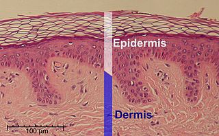
<span class="mw-page-title-main">Epidermis</span> Outermost of the three layers that make up the skin

The epidermis is the outermost of the three layers that comprise the skin, the inner layers being the dermis and hypodermis. The epidermis layer provides a barrier to infection from environmental pathogens and regulates the amount of water released from the body into the atmosphere through transepidermal water loss.

<span class="mw-page-title-main">Keratin 16</span> Protein-coding gene in the species Homo sapiens

Keratin 16 is a protein that in humans is encoded by the KRT16 gene.

<span class="mw-page-title-main">Atopy</span> Predisposition towards allergy

Atopy is the tendency to produce an exaggerated immunoglobulin E (IgE) immune response to otherwise harmless substances in the environment. Allergic diseases are clinical manifestations of such inappropriate, atopic responses.

<span class="mw-page-title-main">Atopic dermatitis</span> Long-term form of skin inflammation

Atopic dermatitis (AD), also known as atopic eczema, is a long-term type of inflammation of the skin. AD is also often called simply eczema but the same term is also used to refer to dermatitis, the larger group of skin conditions. AD results in itchy, red, swollen, and cracked skin. Clear fluid may come from the affected areas, which can thicken over time.

<span class="mw-page-title-main">Urocanic acid</span> Chemical compound

Urocanic acid is an intermediate in the catabolism of L-histidine. The cis-urocanic acid isomer is rare.
<span class="mw-page-title-main">Interleukin 33</span> IL-33 induces helper T cells, mast cells, eosinophils and basophils to produce type 2 cytokines.

Interleukin 33 (IL-33) is a protein that in humans is encoded by the IL33 gene.

<span class="mw-page-title-main">Histidine ammonia-lyase</span>

Histidine ammonia-lyase is an enzyme that in humans is encoded by the HAL gene. It converts histidine into ammonia and urocanic acid. Its systematic name is L-histidine ammonia-lyase (urocanate-forming).

Corneocytes are terminally differentiated keratinocytes and compose most of the stratum corneum, the outermost layer of the epidermis. They are regularly replaced through desquamation and renewal from lower epidermal layers and are essential for its function as a skin barrier.

<span class="mw-page-title-main">Neuropeptide S receptor</span> Protein-coding gene in the species Homo sapiens

The neuropeptide S receptor (NPSR) is a member of the G-protein coupled receptor superfamily of integral membrane proteins which binds neuropeptide S (NPS). It was formerly an orphan receptor, GPR154, until the discovery of neuropeptide S as the endogenous ligand. Increased expression of this gene in ciliated cells of the respiratory epithelium and in bronchial smooth muscle cells is associated with asthma. This gene is a member of the G protein-coupled receptor 1 family and encodes a plasma membrane protein. Mutations in this gene have also been associated with this disease.

Keratohyalin is a protein structure found in cytoplasmic granules of the keratinocytes in the stratum granulosum of the epidermis. Keratohyalin granules (KHG) mainly consist of keratin, profilaggrin, loricrin and trichohyalin proteins which contribute to cornification or keratinization, the process of the formation of epidermal cornified cell envelope. During the keratinocyte differentiation, these granules maturate and expand in size, which leads to the conversion of keratin tonofilaments into a homogenous keratin matrix, an important step in cornification.
<span class="mw-page-title-main">KLK7</span> Protein-coding gene in the species Homo sapiens

Kallikrein-related peptidase 7 (KLK7) is a serine protease that in humans is encoded by the KLK7 gene. KLK7 was initially purified from the epidermis and characterised as stratum corneum chymotryptic enzyme (SCCE). It was later identified as the seventh member of the human kallikrein family, which includes fifteen homologous serine proteases located on chromosome 19 (19q13).
<span class="mw-page-title-main">Thymic stromal lymphopoietin</span> Cytokine, alarmin, and growth factor.

Thymic stromal lymphopoietin (TSLP) is an interleukin (IL)-2-like cytokine, alarmin, and growth factor involved in numerous physiological and pathological processes, primarily those of the immune system. It shares a common ancestor with IL-7.

<span class="mw-page-title-main">Peeling skin syndrome</span> Medical condition

Peeling skin syndrome is an autosomal recessive disorder characterized by lifelong peeling of the stratum corneum, and may be associated with pruritus, short stature, and easily removed anagen hair.

TGM5 is a transglutaminase enzyme.

<span class="mw-page-title-main">Irwin McLean</span> Emeritus Professor of Genetic Medicine

William Henry Irwin McLean is an Irish geneticist who is emeritus professor of genetic medicine, at the School of Life Sciences, University of Dundee.

Mast cell activation syndrome (MCAS) is a term referring to one of two types of mast cell activation disorder (MCAD); the other type is idiopathic MCAD. MCAS is an immunological condition in which mast cells, a type of white blood cell, inappropriately and excessively release chemical mediators, such as histamine, resulting in a range of chronic symptoms, sometimes including anaphylaxis or near-anaphylaxis attacks. Primary symptoms include cardiovascular, dermatological, gastrointestinal, neurological, and respiratory problems.

The epidermal differentiation complex (EDC) is a gene complex comprising over fifty genes encoding proteins involved in the terminal differentiation and cornification of keratinocytes, the primary cell type of the epidermis. In humans, the complex is located on a 1.9 Mbp stretch within chromosome 1q21. The proteins encoded by EDC genes are closely related in terms of function, and evolutionarily they belong to three distinct gene families: the cornified envelope precursor family, the S100 protein family and the S100 fused type protein (SFTP) family.

<span class="mw-page-title-main">Ormdl sphingolipid biosynthesis regulator 3</span> Protein-coding gene in the species Homo sapiens

ORMDL sphingolipid biosynthesis regulator 3 is a protein that in humans is encoded by the ORMDL3 gene. Variants affecting the expression of this gene are associated with asthma in childhood. Transgenic mice which overexpress human ORMDL3 have increased levels of IgE. This correlated with increased numbers of macrophages, neutrophils, eosinophils, CD4+ and enhanced Th2 cytokine levels in the lung tissue.

References

  1. 1 2 3 GRCh38: Ensembl release 89: ENSG00000143631 Ensembl, May 2017
  2. "Human PubMed Reference:". National Center for Biotechnology Information, U.S. National Library of Medicine.
  3. Markova NG, Marekov LN, Chipev CC, Gan SQ, Idler WW, Steinert PM (January 1993). "Profilaggrin is a major epidermal calcium-binding protein". Molecular and Cellular Biology. 13 (1): 613–25. doi:10.1128/MCB.13.1.613. PMC   358940 . PMID   8417356.
  4. Kypriotou M, Huber M, Hohl D (September 2012). "The human epidermal differentiation complex: cornified envelope precursors, S100 proteins and the 'fused genes' family". Experimental Dermatology. 21 (9): 643–9. doi:10.1111/j.1600-0625.2012.01472.x. PMID   22507538. S2CID   5435031.
  5. Steinbinder, Julia; Sachslehner, Attila Placido; Holthaus, Karin Brigit; Eckhart, Leopold (2024-04-23). "Comparative genomics of sirenians reveals evolution of filaggrin and caspase-14 upon adaptation of the epidermis to aquatic life". Scientific Reports. 14 (1): 9278. doi:10.1038/s41598-024-60099-2. ISSN   2045-2322. PMC   11039687 . PMID   38653760.
  6. 1 2 Ovaere P, Lippens S, Vandenabeele P, Declercq W (September 2009). "The emerging roles of serine protease cascades in the epidermis". Trends in Biochemical Sciences. 34 (9): 453–63. doi:10.1016/j.tibs.2009.08.001. PMID   19726197.
  7. Harding CR, Scott IR (November 1983). "Histidine-rich proteins (filaggrins): structural and functional heterogeneity during epidermal differentiation". Journal of Molecular Biology. 170 (3): 651–73. doi:10.1016/s0022-2836(83)80126-0. PMID   6195345.
  8. Jungersted JM, Scheer H, Mempel M, Baurecht H, Cifuentes L, Høgh JK, Hellgren LI, Jemec GB, Agner T, Weidinger S (July 2010). "Stratum corneum lipids, skin barrier function and filaggrin mutations in patients with atopic eczema". Allergy. 65 (7): 911–8. doi:10.1111/j.1398-9995.2010.02326.x. PMID   20132155. S2CID   24679127.
  9. Fluhr JW, Elias PM, Man MQ, Hupe M, Selden C, Sundberg JP, Tschachler E, Eckhart L, Mauro TM, Feingold KR (August 2010). "Is the filaggrin-histidine-urocanic acid pathway essential for stratum corneum acidification?". The Journal of Investigative Dermatology. 130 (8): 2141–4. doi:10.1038/jid.2010.74. PMC   4548931 . PMID   20376063.
  10. 1 2 Palmer CN, Irvine AD, Terron-Kwiatkowski A, Zhao Y, Liao H, Lee SP, Goudie DR, Sandilands A, Campbell LE, Smith FJ, O'Regan GM, Watson RM, Cecil JE, Bale SJ, Compton JG, DiGiovanna JJ, Fleckman P, Lewis-Jones S, Arseculeratne G, Sergeant A, Munro CS, El Houate B, McElreavey K, Halkjaer LB, Bisgaard H, Mukhopadhyay S, McLean WH (April 2006). "Common loss-of-function variants of the epidermal barrier protein filaggrin are a major predisposing factor for atopic dermatitis". Nature Genetics. 38 (4): 441–6. doi:10.1038/ng1767. PMID   16550169. S2CID   2500278.
  11. Weidinger S, Illig T, Baurecht H, Irvine AD, Rodriguez E, Diaz-Lacava A, Klopp N, Wagenpfeil S, Zhao Y, Liao H, Lee SP, Palmer CN, Jenneck C, Maintz L, Hagemann T, Behrendt H, Ring J, Nothen MM, McLean WH, Novak N (July 2006). "Loss-of-function variations within the filaggrin gene predispose for atopic dermatitis with allergic sensitizations". The Journal of Allergy and Clinical Immunology. 118 (1): 214–9. doi:10.1016/j.jaci.2006.05.004. PMID   16815158.
  12. Nomura T, Sandilands A, Akiyama M, Liao H, Evans AT, Sakai K, Ota M, Sugiura H, Yamamoto K, Sato H, Palmer CN, Smith FJ, McLean WH, Shimizu H (February 2007). "Unique mutations in the filaggrin gene in Japanese patients with ichthyosis vulgaris and atopic dermatitis". The Journal of Allergy and Clinical Immunology. 119 (2): 434–40. doi:10.1016/j.jaci.2006.12.646. PMID   17291859.
  13. Schellekens GA, de Jong BA, van den Hoogen FH, van de Putte LB, van Venrooij WJ (January 1998). "Citrulline is an essential constituent of antigenic determinants recognized by rheumatoid arthritis-specific autoantibodies". The Journal of Clinical Investigation. 101 (1): 273–81. doi:10.1172/JCI1316. PMC   508564 . PMID   9421490.
  14. Basu K, Palmer CN, Lipworth BJ, McLean WH, Terron-Kwiatkowski A, Zhao Y, Liao H, Smith FJ, Mitra A, Mukhopadhyay S (September 2008). "Filaggrin null mutations are associated with increased asthma exacerbations in children and young adults" (PDF). Allergy. 63 (9): 1211–7. doi:10.1111/j.1398-9995.2008.01660.x. PMID   18307574. S2CID   8105444.
  15. Palmer CN, Ismail T, Lee SP, Terron-Kwiatkowski A, Zhao Y, Liao H, Smith FJ, McLean WH, Mukhopadhyay S (July 2007). "Filaggrin null mutations are associated with increased asthma severity in children and young adults". The Journal of Allergy and Clinical Immunology. 120 (1): 64–8. doi: 10.1016/j.jaci.2007.04.001 . PMID   17531295.
  16. Henderson J, Northstone K, Lee SP, Liao H, Zhao Y, Pembrey M, Mukhopadhyay S, Smith GD, Palmer CN, McLean WH, Irvine AD (April 2008). "The burden of disease associated with filaggrin mutations: a population-based, longitudinal birth cohort study". The Journal of Allergy and Clinical Immunology. 121 (4): 872–7.e9. doi: 10.1016/j.jaci.2008.01.026 . PMID   18325573.
  17. Tavendale R, Macgregor DF, Mukhopadhyay S, Palmer CN (April 2008). "A polymorphism controlling ORMDL3 expression is associated with asthma that is poorly controlled by current medications". The Journal of Allergy and Clinical Immunology. 121 (4): 860–3. doi: 10.1016/j.jaci.2008.01.015 . PMID   18395550.
  18. Bisgaard H, Simpson A, Palmer CN, Bønnelykke K, McLean I, Mukhopadhyay S, Pipper CB, Halkjaer LB, Lipworth B, Hankinson J, Woodcock A, Custovic A (June 2008). "Gene-environment interaction in the onset of eczema in infancy: filaggrin loss-of-function mutations enhanced by neonatal cat exposure". PLOS Medicine. 5 (6): e131. doi: 10.1371/journal.pmed.0050131 . PMC   2504043 . PMID   18578563.