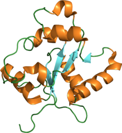
Toll-like receptor 2 also known as TLR2 is a protein that in humans is encoded by the TLR2 gene. [5] TLR2 has also been designated as CD282 (cluster of differentiation 282). TLR2 is one of the toll-like receptors and plays a role in the immune system. TLR2 is a membrane protein, a receptor, which is expressed on the surface of certain cells and recognizes foreign substances and passes on appropriate signals to the cells of the immune system.
The protein encoded by this gene is a member of the toll-like receptor (TLR) family, which plays a fundamental role in pathogen recognition and activation of innate immunity. TLRs are highly conserved from Drosophila to humans and share structural and functional similarities. They recognize pathogen-associated molecular patterns (PAMPs) that are expressed on infectious agents, and mediate the production of cytokines necessary for the development of effective immunity. The various TLRs exhibit different patterns of expression. This gene is expressed most abundantly in peripheral blood leukocytes, and mediates host response to Gram-positive bacteria [6] and yeast via stimulation of NF-κB. [7]
In the intestine, TLR2 regulates the expression of CYP1A1, [8] which is a key enzyme in detoxication of carcinogenic polycyclic aromatic hydrocarbons such as benzo(a)pyrene. [9]
The immune system recognizes foreign pathogens and eliminates them. This occurs in several phases. In the early inflammation phase, pathogens are recognized by antibodies that are already present (innate or acquired through prior infection; see also cross-reactivity). Immune-system components (e.g. complement) are bound to the antibodies and kept near, in reserve to disable them via phagocytosis by scavenger cells (e.g. macrophages). Dendritic cells are likewise capable of phagocytizing but do not do it for the purpose of direct pathogen elimination. Rather, they infiltrate the spleen and lymph nodes, and each presents components of an antigen there, as the result of which specific antibodies are formed that recognize precisely that antigen.
These newly formed antibodies would arrive too late in an acute infection, however, so what we think of as "immunology" constitutes only the second half of the process. Because this phase would always start too late to play an essential role in the defense process, a faster-acting principle is applied ahead of it, one that occurs only in forms of life that are phylogenetically more highly developed.
Pattern-recognition receptors (PRRs) come into play here. These are receptors that recognize the gross, primarily structural features of molecules not innate to the host organism. PRRs include, for example, lipids with a totally different basic chemical structure. Such receptors are bound directly to cells of the immune system and cause immediate activation of their respective nonspecific immune cells.
A prime example of such a foreign ligand is bacterial endotoxin. When endotoxin enters the bloodstream, it activates the early-phase response, which can lead to septic shock and disseminated intravascular coagulation.
As a membrane surface receptor, TLR2 recognizes many bacterial, fungal, viral, and certain endogenous substances. In general, this results in the uptake (internalization, phagocytosis) of bound molecules by endosomes/phagosomes and in cellular activation; thus such elements of innate immunity as macrophages, PMNs and dendritic cells assume functions of nonspecific immune defense, B1a and MZ B cells form the first antibodies, and specific antibody formation gets started in the process. Cytokines participating in this include tumor necrosis factor-alpha (TNF-α) and various interleukins (IL-1α, IL-1β, IL-6, IL-8, IL-12). Before the TLRs were known, several of the substances mentioned were classified as modulins. Due to the cytokine pattern, which corresponds more closely to Th1, an immune deviation is seen in this direction in most experimental models, away from Th2 characteristics. Conjugates are being developed as vaccines or are already being used without a priori knowledge.
A peculiarity first recognized in 2006 is the expression of TLR2 on Tregs (a type of T cell), which experience both TCR-controlled proliferation and functional inactivation. This leads to disinhibition of the early inflammation phase and of specific antibody formation. Following a reduction in pathogen count, many pathogen-specific Tregs are present that, now without a TLR2 signal, become active and inhibit the specific and inflammatory immune reactions (see also TNF-β, IL-10). Older literature that ascribes a direct immunity-stimulating effect via TLR2 to a given molecule must be interpreted in light of the fact that the TLR2 knockouts employed typically have very few Tregs.
Functionally relevant polymorphisms are reported that cause functional impairment and thus, in general, reduced survival rates, in particular in infections/sepsis with Gram-positive bacteria.
Signal transduction is depicted under Toll-like receptor.
TLR2 is expressed on microglia, Schwann cells, monocytes, macrophages, dendritic cells, polymorphonuclear leukocytes (PMNs or PMLs), B cells (B1a, MZ B, B2), and T cells, including Tregs (CD4+CD25+ regulatory T cells). In some cases, it occurs in a heterodimer (combination molecule), e.g., paired with TLR-1 or TLR-6. TLR2 is also found in the epithelia of air passages, pulmonary alveoli, renal tubules, and the Bowman's capsules in renal corpuscles. TLR2 is also expressed by intestinal epithelial cells and subsets of lamina propria mononuclear cells in the gastrointestinal tract. [10] In the skin, it is found on keratinocytes and sebaceous glands; spc1 is induced here, allowing a bactericidal sebum to be formed.
TLR2 gene has been observed progressively downregulated in Human papillomavirus-positive neoplastic keratinocytes derived from uterine cervical preneoplastic lesions at different levels of malignancy. [11] For this reason, TLR2 is likely to be associated with tumorigenesis and may be a potential prognostic marker for uterine cervical preneoplastic lesions progression. [11]
The following ligands have been reported to be agonists of the toll-like receptor 2:
Agonist | Organism |
---|---|
Lipoteichoic acid | Gram-positive bacteria |
atypical LPS | Leptospirosis and Porphyromonas gingivalis |
MALP-2 and MALP-404 (lipoproteins) | Mycoplasma |
- | Chlamydophila pneumoniae |
OspA | Borrelia burgdorferi (Lyme disease) |
Porin | Neisseria meningitidis , Haemophilus influenzae |
Antigen mixtures | Cutibacterium acnes |
LcrV | Yersinia |
Lipomannan | Mycobacterium : Mycobacterium tuberculosis |
GPI anchor | Trypanosoma cruzi |
Lysophosphatidylserine | Schistosoma mansoni |
Lipophosphoglycan (LPG) | Leishmania major |
Glycophosphatidylinositol (GPI) | Plasmodium falciparum |
Zymosan (a beta-glucan) [12] [13] | Saccharomyces cerevisiae |
- | Malassezia (commensal yeast) |
Antigen mixtures | Aspergillus fumigatus , Candida albicans |
hsp60, as peptide transporter and adjuvant for antigen presentation | - |
Glycoprotein (gH/gL, gB) [14] | Herpes simplex virus |
- | Varicella zoster virus |
- | Cytomegalovirus (CMV) |
Hemagglutinin | Measles |
Active hexose correlated compound (AHCC) [15] | Shiitake |
TLR 2 has been shown to interact with TLR 1 [16] and TOLLIP. [17] It has been shown that TLR2 can interact with spike and E-protein of SARS-CoV-2. [18] The result of these interactions can be the activation of the immune system. [18]
TLR2 resides on the plasma membrane where it responds to lipid-containing PAMPs such as lipoteichoic acid and di- and tri-acylated cysteine-containing lipopeptides. It does this by forming dimeric complexes with either TLR 1 or TLR6 on the plasma membrane. [19] TLR2 interactions with malarial glycophosphatidylinositols of Plasmodium falciparum was shown [20] and a detailed structure of TLR–GPI interactions was computationally predicted. [21]
Various single nucleotide polymorphisms (SNPs) of the TLR2 have been identified [22] and for some of them an association with faster progression and a more severe course of sepsis in critically ill patients was reported. [23] No association with occurrence of severe staphylococcal infection was found. [24] Moreover, a recent study reported rs111200466, a TLR2 promoter insertion/deletion polymorphism as a prognosis factor in HIV-1 disease progression. The authors showed a correlation of the polymorphism with a faster progression to the CD4+ < 200 cells/μL outcome for the deletion allele carriers. [25]
The immune system is a network of biological systems that protects an organism from diseases. It detects and responds to a wide variety of pathogens, from viruses to bacteria, as well as cancer cells, parasitic worms, and also objects such as wood splinters, distinguishing them from the organism's own healthy tissue. Many species have two major subsystems of the immune system. The innate immune system provides a preconfigured response to broad groups of situations and stimuli. The adaptive immune system provides a tailored response to each stimulus by learning to recognize molecules it has previously encountered. Both use molecules and cells to perform their functions.
Natural killer cells, also known as NK cells, are a type of cytotoxic lymphocyte critical to the innate immune system. They are a kind of large granular lymphocytes (LGL), and belong to the rapidly expanding family of known innate lymphoid cells (ILC) and represent 5–20% of all circulating lymphocytes in humans. The role of NK cells is analogous to that of cytotoxic T cells in the vertebrate adaptive immune response. NK cells provide rapid responses to virus-infected cells, stressed cells, tumor cells, and other intracellular pathogens based on signals from several activating and inhibitory receptors. Most immune cells detect the antigen presented on major histocompatibility complex I (MHC-I) on infected cell surfaces, but NK cells can recognize and kill stressed cells in the absence of antibodies and MHC, allowing for a much faster immune reaction. They were named "natural killers" because of the notion that they do not require activation to kill cells that are missing "self" markers of MHC class I. This role is especially important because harmful cells that are missing MHC I markers cannot be detected and destroyed by other immune cells, such as T lymphocyte cells.
Toll-like receptors (TLRs) are a class of proteins that play a key role in the innate immune system. They are single-spanning receptors usually expressed on sentinel cells such as macrophages and dendritic cells, that recognize structurally conserved molecules derived from microbes. Once these microbes have reached physical barriers such as the skin or intestinal tract mucosa, they are recognized by TLRs, which activate immune cell responses. The TLRs include TLR1, TLR2, TLR3, TLR4, TLR5, TLR6, TLR7, TLR8, TLR9, TLR10, TLR11, TLR12, and TLR13. Humans lack genes for TLR11, TLR12 and TLR13 and mice lack a functional gene for TLR10. The receptors TLR1, TLR2, TLR4, TLR5, TLR6, and TLR10 are located on the cell membrane, whereas TLR3, TLR7, TLR8, and TLR9 are located in intracellular vesicles.
Pathogen-associated molecular patterns (PAMPs) are small molecular motifs conserved within a class of microbes, but not present in the host. They are recognized by toll-like receptors (TLRs) and other pattern recognition receptors (PRRs) in both plants and animals. This allows the innate immune system to recognize pathogens and thus, protect the host from infection.
Pattern recognition receptors (PRRs) play a crucial role in the proper function of the innate immune system. PRRs are germline-encoded host sensors, which detect molecules typical for the pathogens. They are proteins expressed mainly by cells of the innate immune system, such as dendritic cells, macrophages, monocytes, neutrophils, as well as by epithelial cells, to identify two classes of molecules: pathogen-associated molecular patterns (PAMPs), which are associated with microbial pathogens, and damage-associated molecular patterns (DAMPs), which are associated with components of host's cells that are released during cell damage or death. They are also called primitive pattern recognition receptors because they evolved before other parts of the immune system, particularly before adaptive immunity. PRRs also mediate the initiation of antigen-specific adaptive immune response and release of inflammatory cytokines.
Gut-associated lymphoid tissue (GALT) is a component of the mucosa-associated lymphoid tissue (MALT) which works in the immune system to protect the body from invasion in the gut.
The innate immune system or nonspecific immune system is one of the two main immunity strategies in vertebrates. The innate immune system is an alternate defense strategy and is the dominant immune system response found in plants, fungi, prokaryotes, and invertebrates.
CD14 is a human protein made mostly by macrophages as part of the innate immune system. It helps to detect bacteria in the body by binding lipopolysaccharide (LPS), a pathogen-associated molecular pattern (PAMP).
Myeloid differentiation primary response 88 (MYD88) is a protein that, in humans, is encoded by the MYD88 gene. originally discovered in the laboratory of Dan A. Liebermann as a Myeloid differentiation primary response gene.
Toll-like receptor 1 (TLR1) is a member of the toll-like receptor (TLR) family of pattern recognition receptors (PRRs) that form the cornerstone of the innate immune system. TLR1 recognizes bacterial lipoproteins and glycolipids in complex with TLR2. TLR1 is a cell surface receptor. In humans, TLR1 is encoded by the TLR1 gene, which is located on chromosome 4.
Toll-like receptor 5, also known as TLR5, is a protein which in humans is encoded by the TLR5 gene. It is a member of the toll-like receptor (TLR) family. TLR5 is known to recognize bacterial flagellin from invading mobile bacteria. It has been shown to be involved in the onset of many diseases, including Inflammatory bowel disease due to the high expression of TLR in intestinal lamina propria dendritic cells. Recent studies have also shown that malfunctioning of TLR5 is likely related to rheumatoid arthritis, osteoclastogenesis, and bone loss. Abnormal TLR5 functioning is related to the onset of gastric, cervical, endometrial and ovarian cancers.
Toll-like receptor 4 (TLR4), also designated as CD284, is a key activator of the innate immune response and plays a central role in the fight against bacterial infections. TLR4 is a transmembrane protein of approximately 95 kDa that is encoded by the TLR4 gene.
Toll-like receptor 6 is a protein that in humans is encoded by the TLR6 gene. TLR6 is a transmembrane protein, member of toll-like receptor family, which belongs to the pattern recognition receptor (PRR) family. TLR6 acts in a heterodimer form with toll-like receptor 2 (TLR2). Its ligands include multiple diacyl lipopeptides derived from gram-positive bacteria and mycoplasma and several fungal cell wall saccharides. After dimerizing with TLR2, the NF-κB intracellular signalling pathway is activated, leading to a pro-inflammatory cytokine production and activation of innate immune response. TLR6 has also been designated as CD286.
Toll-like receptor 10 is a protein that in humans is encoded by the TLR10 gene. TLR10 has also been designated as CD290 . TLR10 has not been extensively studied because it is a pseudogene in mice, though all other mammalian species contain an intact copy of the TLR10 gene. Unlike other TLRs, TLR10 does not activate the immune system and has instead been shown to suppress inflammatory signaling on primary human cells. This makes TLR10 unique among the TLR family. TLR10 was thought to be an "orphan" receptor, however, recent studies have identified ligands for TLR10 and these include HIV-gp41. Ligands for TLR2 are potential ligands for TLR10.
Toll interacting protein, also known as TOLLIP, is an inhibitory adaptor protein that in humans is encoded by the TOLLIP gene.
Macrophage receptor with collagenous structure (MARCO) is a protein that in humans is encoded by the MARCO gene. MARCO is a class A scavenger receptor that is found on particular subsets of macrophages. Scavenger receptors are pattern recognition receptors (PRRs) found most commonly on immune cells. Their defining feature is that they bind to polyanions and modified forms of a type of cholesterol called low-density lipoprotein (LDL). MARCO is able to bind and phagocytose these ligands and pathogen-associated molecular patterns (PAMPs), leading to the clearance of pathogens and cell signaling events that lead to inflammation. As part of the innate immune system, MARCO clears, or scavenges, pathogens, which leads to inflammatory responses. The scavenger receptor cysteine-rich (SRCR) domain at the end of the extracellular side of MARCO binds ligands to activate the subsequent immune responses. MARCO expression on macrophages has been associated with tumor development and also with Alzheimer's disease, via decreased responses of cells when ligands bind to MARCO.
Damage-associated molecular patterns (DAMPs) are molecules within cells that are a component of the innate immune response released from damaged or dying cells due to trauma or an infection by a pathogen. They are also known as danger signals, and alarmins because they serve as warning signs to alert the organism to any damage or infection to its cells. DAMPs are endogenous danger signals that are discharged to the extracellular space in response to damage to the cell from mechanical trauma or a pathogen. Once a DAMP is released from the cell, it promotes a noninfectious inflammatory response by binding to a pattern recognition receptor (PRR). Inflammation is a key aspect of the innate immune response; it is used to help mitigate future damage to the organism by removing harmful invaders from the affected area and start the healing process. As an example, the cytokine IL-1α is a DAMP that originates within the nucleus of the cell which, once released to the extracellular space, binds to the PRR IL-1R, which in turn initiates an inflammatory response to the trauma or pathogen that initiated the release of IL-1α. In contrast to the noninfectious inflammatory response produced by DAMPs, pathogen-associated molecular patterns (PAMPs) initiate and perpetuate the infectious pathogen-induced inflammatory response. Many DAMPs are nuclear or cytosolic proteins with defined intracellular function that are released outside the cell following tissue injury. This displacement from the intracellular space to the extracellular space moves the DAMPs from a reducing to an oxidizing environment, causing their functional denaturation, resulting in their loss of function. Outside of the aforementioned nuclear and cytosolic DAMPs, there are other DAMPs originated from different sources, such as mitochondria, granules, the extracellular matrix, the endoplasmic reticulum, and the plasma membrane.
Long-term close-knit interactions between symbiotic microbes and their host can alter host immune system responses to other microorganisms, including pathogens, and are required to maintain proper homeostasis. The immune system is a host defense system consisting of anatomical physical barriers as well as physiological and cellular responses, which protect the host against harmful microorganisms while limiting host responses to harmless symbionts. Humans are home to 1013 to 1014 bacteria, roughly equivalent to the number of human cells, and while these bacteria can be pathogenic to their host most of them are mutually beneficial to both the host and bacteria.
Toll-like receptor 11 (TLR11) is a protein that in mice and rats is encoded by the gene TLR11, whereas in humans it is represented by a pseudogene. TLR11 belongs to the toll-like receptor (TLR) family and the interleukin-1 receptor/toll-like receptor superfamily. In mice, TLR11 has been shown to recognise (bacterial) flagellin and (eukaryotic) profilin present on certain microbes, it helps propagate a host immune response. TLR11 plays a fundamental role in both the innate and adaptive immune responses, through the activation of tumor necrosis factor-alpha, the interleukin 12 (IL-12) response, and interferon-gamma (IFN-gamma) secretion. TLR11 mounts an immune response to multiple microbes, including Toxoplasma gondii, Salmonella species, and uropathogenic E. coli, and likely many other species due to the highly conserved nature of flagellin and profilin.
Mucosal immunology is the study of immune system responses that occur at mucosal membranes of the intestines, the urogenital tract, and the respiratory system. The mucous membranes are in constant contact with microorganisms, food, and inhaled antigens. In healthy states, the mucosal immune system protects the organism against infectious pathogens and maintains a tolerance towards non-harmful commensal microbes and benign environmental substances. Disruption of this balance between tolerance and deprivation of pathogens can lead to pathological conditions such as food allergies, irritable bowel syndrome, susceptibility to infections, and more.
This article incorporates text from the United States National Library of Medicine, which is in the public domain.