PRDM16

Last updated
PRDM16
PRDM16.png
Available structures
PDB Ortholog search: PDBe RCSB
Identifiers
Aliases PRDM16 , CMD1LL, LVNC8, MEL1, PFM13, PR domain 16, PR/SET domain 16, KMT8F
External IDs OMIM: 605557 MGI: 1917923 HomoloGene: 11139 GeneCards: PRDM16
Orthologs
SpeciesHumanMouse
Entrez
Ensembl
UniProt
RefSeq (mRNA)

NM_022114
NM_199454

NM_001177995
NM_001291026
NM_001291029
NM_027504

RefSeq (protein)

NP_071397
NP_955533

NP_001171466
NP_001277955
NP_001277958
NP_081780

Location (UCSC) Chr 1: 3.07 – 3.44 Mb Chr 4: 154.4 – 154.72 Mb
PubMed search [3] [4]
Wikidata
View/Edit Human View/Edit Mouse

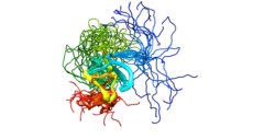
PR domain containing 16, also known as PRDM16, is a protein which in humans is encoded by the PRDM16 gene. [5] [6]

Contents

PRDM16 acts as a transcription coregulator that controls the development of brown adipocytes in brown adipose tissue. [7] Previously, this coregulator was believed to be present only in brown adipose tissue, but more recent studies have shown that PRDM16 is highly expressed in subcutaneous white adipose tissue as well. [7]

Function

The protein encoded by this gene is a zinc finger transcription factor. [6] PRDM16 controls the cell fate between muscle and brown fat cells. Loss of PRDM16 from brown fat precursors causes a loss of brown fat characteristics and promotes muscle differentiation. [8]

Clinical significance

The reciprocal translocation t(1;3)(p36;q21) occurs in a subset of myelodysplastic syndrome (MDS) and acute myeloid leukemia (AML). This gene is located near the 1p36.3 breakpoint and has been shown to be specifically expressed in the t(1:3)(p36;q21)-positive MDS/AML. The protein encoded by this gene contains an N-terminal PR domain. The translocation results in the overexpression of a truncated version of this protein that lacks the PR domain, which may play an important role in the pathogenesis of MDS and AML. Alternatively spliced transcript variants encoding distinct isoforms have been reported. [6]

PRDM16 in BAT

Brown adipose tissue (BAT) oxidizes chemical energy to produce heat. This heat energy can act as a defense against hypothermia and obesity. [7] PRDM16 is highly enriched in brown adipose cells as compared to white adipose cells, and plays a role in these thermogenic processes in brown adipose tissue. PRDM16 activates brown fat cell identity and can control the determination of brown adipose fate. A knock-out of PRDM16 in mice shows a loss of brown cell characteristics, showing that PRDM16 activity is important in determining brown adipose fate. [9] Brown adipocytes consist of densely packed mitochondria that contain uncoupling protein 1 (UCP-1). UCP-1 plays a key role in brown adipocyte thermogenesis. The presence of PRDM16 in adipose tissue causes a significant up-regulation of thermogenic genes, such as UCP-1 and CIDEA, resulting in thermogenic heat production. [7] Understanding and stimulating the thermogenic processes in brown adipocytes provides possible therapeutic options for treating obesity. [9]

PRDM16 in WAT

White adipose tissue (WAT) primarily stores excess energy in the form of triglycerides. [7] [9] Recent research has shown that PRDM16 is present in subcutaneous white adipose tissue. [7] The activity of PRDM16 in white adipose tissue leads to the production of brown fat-like adipocytes within white adipose tissue, called beige cells (also called brite cells). These beige cells have a brown adipose tissue-like phenotype and actions, including thermogenic processes seen in BAT. [7] In mice, the levels of PRDM16 within WAT, specifically anterior subcutaneous WAT and inguinal subcutaneous WAT, is about 50% that of interscapular BAT, both in protein expression and in mRNA quantity. [7] This expression takes place primarily within mature adipocytes. Transgenic aP2-PRDM16 mice were used in a study to observe the effects of PRDM16 expression in WAT. [7] The study found that the presence of PRDM16 in subcutaneous WAT leads to a significant up-regulation of brown-fat selective genes UCP-1, CIDEA, and PPARGC1A. This up-regulation lead to the development of a BAT-like phenotype within the white adipose tissue. Expression of PRDM16 has also been shown to protect against high-fat diet induced weight gain. [7] Seale et al.’s experiment with aP2-PRDM16 transgenic mice and wild type mice showed that transgenic mice eating a 60% high-fat diet had significantly less weight gain than wild type mice on the same diet. Seale et al. determined the weight difference was not due to differences in food intake, as both transgenic and wild type mice were consuming the same amount of food on a daily basis. Rather, the weight difference stemmed from higher energy expenditure in the transgenic mice. Another of Seale et al.’s experiments showed the transgenic mice consumed a greater volume of oxygen over a 72-hour period than the wild type mice, showing a greater amount of energy expenditure in the transgenic mice. [7] This energy expenditure in turn is attributed to PRDM16’s ability to up-regulate UCP-1 and CIDEA gene expression, resulting in thermogenesis.

If human WAT expresses PRDM16 as in mice, this WAT could be a potential target for stimulating energy expenditure and combating obesity.

Notes

  1. 1 2 3 GRCh38: Ensembl release 89: ENSG00000142611 - Ensembl, May 2017
  2. 1 2 3 GRCm38: Ensembl release 89: ENSMUSG00000039410 - Ensembl, May 2017
  3. "Human PubMed Reference:". National Center for Biotechnology Information, U.S. National Library of Medicine.
  4. "Mouse PubMed Reference:". National Center for Biotechnology Information, U.S. National Library of Medicine.
  5. Mochizuki N, Shimizu S, Nagasawa T, Tanaka H, Taniwaki M, Yokota J, Morishita K (November 2000). "A novel gene, MEL1, mapped to 1p36.3 is highly homologous to the MDS1/EVI1 gene and is transcriptionally activated in t(1;3)(p36;q21)-positive leukemia cells". Blood. 96 (9): 3209–14. doi:10.1182/blood.V96.9.3209. PMID   11050005.
  6. 1 2 3 "Entrez Gene: PRDM16 PR domain containing 16".
  7. 1 2 3 4 5 6 7 8 9 10 11 Patrick Seale, Heather M. Conroe, Jennifer Estall, Shingo Kajimura, Andrea Frontini, Jeff Ishibashi, Paul Cohen, Saverio Cinti, Bruce M. Spiegelman (January 2011). "Prdm16 determines the thermogenic program of subcutaneous white adipose tissue in mice". The Journal of Clinical Investigation. 121 (1): 96–105. doi:10.1172/JCI44271. PMC   3007155 . PMID   21123942.
  8. Seale P, Bjork B, Yang W, Kajimura S, Chin S, Kuang S, Scimè A, Devarakonda S, Conroe HM, Erdjument-Bromage H, Tempst P, Rudnicki MA, Beier DR, Spiegelman BM (August 2008). "PRDM16 controls a brown fat/skeletal muscle switch". Nature. 454 (7207): 961–7. Bibcode:2008Natur.454..961S. doi:10.1038/nature07182. PMC   2583329 . PMID   18719582.
  9. 1 2 3 Patrick Seale, Shingo Kajimura, Wenli Yang, Sherry Chin, Lindsay Rohas, Marc Uldry, Geneviève Tavernier, Dominique Langin, Bruce M Spiegelman (July 2007). "Transcriptional Control of Brown Fat Determination by PRDM16". Cell Metabolism. 6 (1): 38–54. doi:10.1016/j.cmet.2007.06.001. PMC   2564846 . PMID   17618855.

Related Research Articles

<span class="mw-page-title-main">Lipolysis</span> Metabolism involving breakdown of lipids

Lipolysis is the metabolic pathway through which lipid triglycerides are hydrolyzed into a glycerol and free fatty acids. It is used to mobilize stored energy during fasting or exercise, and usually occurs in fat adipocytes. The most important regulatory hormone in lipolysis is insulin; lipolysis can only occur when insulin action falls to low levels, as occurs during fasting. Other hormones that affect lipolysis include leptin, glucagon, epinephrine, norepinephrine, growth hormone, atrial natriuretic peptide, brain natriuretic peptide, and cortisol.

<span class="mw-page-title-main">Brown adipose tissue</span> Type of adipose tissue

Brown adipose tissue (BAT) or brown fat makes up the adipose organ together with white adipose tissue. Brown adipose tissue is found in almost all mammals.

<span class="mw-page-title-main">Adipose tissue</span> Loose connective tissue composed mostly by adipocytes

Adipose tissue (also known as body fat, or simply fat) is a loose connective tissue composed mostly of adipocytes. In addition to adipocytes, adipose tissue contains the stromal vascular fraction(SVF) of cells including preadipocytes, fibroblasts, vascular endothelial cells and a variety of immune cells such as adipose tissue macrophages. Adipose tissue is derived from preadipocytes. Its main role is to store energy in the form of lipids, although it also cushions and insulates the body. Far from being hormonally inert, adipose tissue has, in recent years, been recognized as a major endocrine organ, as it produces hormones such as leptin, estrogen, resistin, and cytokines (especially TNFα). In obesity, adipose tissue is also implicated in the chronic release of pro-inflammatory markers known as adipokines, which are responsible for the development of metabolic syndrome, a constellation of diseases, including type 2 diabetes, cardiovascular disease and atherosclerosis. The two types of adipose tissue are white adipose tissue (WAT), which stores energy, and brown adipose tissue (BAT), which generates body heat. The formation of adipose tissue appears to be controlled in part by the adipose gene. Adipose tissue – more specifically brown adipose tissue – was first identified by the Swiss naturalist Conrad Gessner in 1551.

<span class="mw-page-title-main">Thermogenin</span> Mammalian protein found in Homo sapiens

Thermogenin is a mitochondrial carrier protein found in brown adipose tissue (BAT). It is used to generate heat by non-shivering thermogenesis, and makes a quantitatively important contribution to countering heat loss in babies which would otherwise occur due to their high surface area-volume ratio.

<span class="mw-page-title-main">Adipocyte</span> Cells that primarily compose adipose tissue, specialized in storing energy as fat

Adipocytes, also known as lipocytes and fat cells, are the cells that primarily compose adipose tissue, specialized in storing energy as fat. Adipocytes are derived from mesenchymal stem cells which give rise to adipocytes through adipogenesis. In cell culture, adipocyte progenitors can also form osteoblasts, myocytes and other cell types.

<span class="mw-page-title-main">Adiponectin</span> Mammalian protein found in Homo sapiens

Adiponectin is a protein hormone and adipokine, which is involved in regulating glucose levels and fatty acid breakdown. In humans, it is encoded by the ADIPOQ gene and is produced primarily in adipose tissue, but also in muscle and even in the brain.

In biochemistry, lipogenesis is the conversion of fatty acids and glycerol into fats, or a metabolic process through which acetyl-CoA is converted to triglyceride for storage in fat. Lipogenesis encompasses both fatty acid and triglyceride synthesis, with the latter being the process by which fatty acids are esterified to glycerol before being packaged into very-low-density lipoprotein (VLDL). Fatty acids are produced in the cytoplasm of cells by repeatedly adding two-carbon units to acetyl-CoA. Triacylglycerol synthesis, on the other hand, occurs in the endoplasmic reticulum membrane of cells by bonding three fatty acid molecules to a glycerol molecule. Both processes take place mainly in liver and adipose tissue. Nevertheless, it also occurs to some extent in other tissues such as the gut and kidney. A review on lipogenesis in the brain was published in 2008 by Lopez and Vidal-Puig. After being packaged into VLDL in the liver, the resulting lipoprotein is then secreted directly into the blood for delivery to peripheral tissues.

<span class="mw-page-title-main">Perilipin-1</span> Protein in humans

Perilipin, also known as lipid droplet-associated protein, perilipin 1, or PLIN, is a protein that, in humans, is encoded by the PLIN gene. The perilipins are a family of proteins that associate with the surface of lipid droplets. Phosphorylation of perilipin is essential for the mobilization of fats in adipose tissue.

<span class="mw-page-title-main">White adipose tissue</span> Fatty tissue composed of white adipocytes

White adipose tissue or white fat is one of the two types of adipose tissue found in mammals. The other kind is brown adipose tissue. White adipose tissue is composed of monolocular adipocytes.

<span class="mw-page-title-main">UCP3</span> Protein-coding gene in the species Homo sapiens

Mitochondrial uncoupling protein 3 is a protein that in humans is encoded by the UCP3 gene. The gene is located in chromosome (11q13.4) with an exon count of 7 and is expressed on the inner mitochondrial membrane. Uncoupling proteins transfer anions from the inner mitochondrial membrane to the outer mitochondrial membrane, thereby separating oxidative phosphorylation from synthesis of ATP, and dissipating energy stored in the mitochondrial membrane potential as heat. Uncoupling proteins also reduce generation of reactive oxygen species.

<span class="mw-page-title-main">GPR3</span> Protein

G-protein coupled receptor 3 is a protein that in humans is encoded by the GPR3 gene. The protein encoded by this gene is a member of the G protein-coupled receptor family of transmembrane receptors and is involved in signal transduction.

<span class="mw-page-title-main">MECOM</span> Protein-coding gene in the species Homo sapiens

MDS1 and EVI1 complex locus protein EVI1 (MECOM) also known as ecotropic virus integration site 1 protein homolog (EVI-1) or positive regulatory domain zinc finger protein 3 (PRDM3) is a protein that in humans is encoded by the MECOM gene. EVI1 was first identified as a common retroviral integration site in AKXD murine myeloid tumors. It has since been identified in a plethora of other organisms, and seems to play a relatively conserved developmental role in embryogenesis. EVI1 is a nuclear transcription factor involved in many signaling pathways for both coexpression and coactivation of cell cycle genes.

<span class="mw-page-title-main">Brain mitochondrial carrier protein 1</span> Protein-coding gene in the species Homo sapiens

Brain mitochondrial carrier protein 1 is a protein that in humans is encoded by the SLC25A14 gene.

<span class="mw-page-title-main">HRASLS3</span> Protein-coding gene in the species Homo sapiens

Group XVI phospholipase A2 also commonly known as adipocyte phospholipase A2 (AdPLA) is an enzyme that in humans is encoded by the PLA2G16 gene. This enzyme has also been identified as PLA2G16, HRASLS3, HREV107, HREV107-3, MGC118754 or H-REV107-1 from studies on class II tumor suppression but not on its enzymatic properties. AdPLA is encoded by a 1.3 kilobase AdPLA messenger RNA and is an 18 kDa protein. It belongs to a superfamily of phospholipase A2 (PLA2) enzymes and is found primarily in adipose tissue. AdPLA regulates adipocyte lipolysis and release of fatty acids through a G-protein coupled pathway involving prostaglandin and EP3. It has also been reported to play a crucial role in the development of obesity in mouse models.

Congenital generalized lipodystrophy is an extremely rare autosomal recessive condition, characterized by an extreme scarcity of fat in the subcutaneous tissues. It is a type of lipodystrophy disorder where the magnitude of fat loss determines the severity of metabolic complications. Only 250 cases of the condition have been reported, and it is estimated that it occurs in 1 in 10 million people worldwide.

<span class="mw-page-title-main">FNDC5</span>

Fibronectin type III domain-containing protein 5, the precursor of irisin, is a type I transmembrane glycoprotein that is encoded by the FNDC5 gene. Irisin is a cleaved version of FNDC5, named after the Greek messenger goddess Iris.

<span class="mw-page-title-main">Adipogenesis</span>

Adipogenesis is the formation of adipocytes from stem cells. It involves 2 phases, determination, and terminal differentiation. Determination is mesenchymal stem cells committing to the adipocyte precursor cells, also known as lipoblasts or preadipocytes which lose the potential to differentiate to other types of cells such as chondrocytes, myocytes, and osteoblasts. Terminal differentiation is that preadipocytes differentiate into mature adipocytes. Adipocytes can arise either from preadipocytes resident in adipose tissue, or from bone-marrow derived progenitor cells that migrate to adipose tissue.

<span class="mw-page-title-main">Bone marrow adipose tissue</span>

Bone marrow adipose tissue (BMAT), sometimes referred to as marrow adipose tissue (MAT), is a type of fat deposit in bone marrow. It increases in states of low bone density -osteoporosis, anorexia nervosa/ caloric restriction, skeletal unweighting such as that which occurs in space travel, and anti-diabetes therapies. BMAT decreases in anaemia, leukaemia, and hypertensive heart failure; in response to hormones such as oestrogen, leptin, and growth hormone; with exercise-induced weight loss or bariatric surgery; in response to chronic cold exposure; and in response to pharmacological agents such as bisphosphonates, teriparatide, and metformin.

<span class="mw-page-title-main">Daniel Ricquier</span> French biochemist

Daniel Ricquier, is a French biochemist known for his work in mitochondria and hereditary metabolic diseases. Ricquier has been a member of the French Academy of Sciences since 2002, and a professor of biochemistry and Molecular Biology at the Faculty of Medicine of the University of Paris Descartes since 2003.

<span class="mw-page-title-main">Barbara Cannon</span> British physiologist (1946-)

Barbara Cannon is a British-Swedish biochemist, physiologist and an academic. She is an emeritus professor at Stockholm University as well as the chairman of the scientific advisory board at The Helmholtz Centre. She is also a consultant at Combigene.

References

Further reading