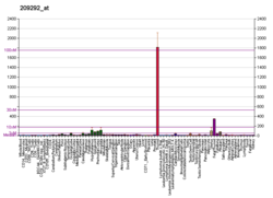
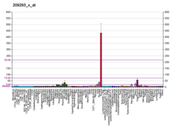
ID4 is a protein coding gene. In humans, it encodes the protein known as DNA-binding protein inhibitor ID-4. [5] [6] This protein is known to be involved in the regulation of many cellular processes during both prenatal development and tumorigenesis. This is inclusive of embryonic cellular growth, senescence, cellular differentiation, apoptosis, and as an oncogene in angiogenesis. [7]
The gene spans 3.3kb on the plus strand. It is composed of 3 exons and during transcription its mRNA is 2343 bp. The encoded protein consists of 161 amino acids, is 16.6 KDa and contains a poly-Ala segment from amino acid 39 to 48, a helix-loop-helix motif from amino acid 65 to 105 and a poly-Pro region from amino acid 118 to 124. This protein is expressed in various tissues. [7]
The ID4 gene is part of the ID gene family. This family is also known as inhibitors of DNA binding protein family and are composed of transcription inhibitory proteins which modulate a number of processes. They are transcriptional regulators that work by negatively regulating their basic helix-loop-helix (bHLH) transcription factors by forming heterodimers. The heterodimer is what inhibits their DNA binding and transcriptional activity.
Transcription factors containing a basic helix-loop-helix (bHLH) motif regulate expression of tissue-specific genes in a number of mammalian and insect systems. DNA-binding activity of the bHLH proteins is dependent on formation of homo- and/or heterodimers. Dominant-negative (antimorph) HLH proteins encoded by Id-related genes, such as ID4, also contain the HLH-dimerization domain but lack the DNA-binding basic domain. Consequently, ID proteins inhibit binding to DNA and transcriptional transactivation by heterodimerization with bHLH proteins. [6]
The ID4 gene plays a pivotal role in development and is a key player in many pathways of embryogenesis and foetal development. ID4 expression is upregulated in embryogenesis during days 9.5 and 13.5 of gestation [8] and restricted to specific cells of the central and peripheral nervous system. [9] ID4 transcription control has both negative and positive regulatory elements inclusive of novel inhibitory functions. [10]
ID4 expression has been shown to be discrete in the early stages, with transcription transiently expressed in subsets of migrating neural crest cells, the dorsal myocardium, the segmental plate mesoderm, and the tail bud. Later stages show ID4 expression in the telencephalic vesicles and corneal epithelium. [11] ID4 expression is only detected in neuronal tissues and the ventral portion of the epithelium in the developing stomach during embryogenesis. [12]
ID4 is expressed in the central nervous system and is required for G1-S transition and to enhance proliferation in early cortical progenitors. It is complexly involved in regulating neural stem cell proliferation and differentiation by inhibiting proliferation of differentiating neurons through enhancement of RB1-mediated pathways. This is either by direct interaction or through interaction with other molecules of the cell cycle machinery. [13] ID4 also regulates the lateral expansion of the proliferative zone in the developing cortex and hippocampus. This is integral to normal brain size formation. ID4 regulates neural progenitor proliferation and differentiation. [13] Its expression is seen in the neural tube much later than other ID genes. [11]
ID4 was also shown to be involved in the regulation of cardiac mesoderm function in frog embryos and human embryonic stem cells. Ablation of the ID gene family mouse embryos showed failure of anterior cardiac progenitor specification and the development of heartless embryos. This study also demonstrated that ID4 protein is involved in the regulating cardiac cell fate by a pathway which represses two inhibitors of cardiogenic mesoderm formation (TCF3 and FOXA2) whilst activating inducers (EVX1, GRRP1, and MESP1). [14]
ID4 has been linked to the molecular pathogenicity of endometriosis. These pathways are still poorly understood. It is thought that ID4 plays a role in the transcription of HOXA9 and CDKN1A which are known to be associated with endometriosis.
A genome wide association study revealed over 100 candidate genes associated with endometriosis. Of these, six were shown to have a highly reliable association, of which the ID4 gene was identified. This is thought to be due to an independent single nucleotide polymorphism at loci rs7739264 near ID4 on chromosome 6p22.3. ID4 is implicated in the molecular pathogenicity of endometriosis as being differentially expressed between the proliferative, early and mid-secretory phases. [15]
ID4 is not expressed in normal ovary and fallopian tubes. It has been shown to be overexpressed in most primary ovarian cancers. The ID4 gene is also seen to be overexpressed in most ovarian, endometrial and breast cancer cell lines. [16] The mechanism behind this is believed to be that ID4 regulates HOXA9 and CDKN1A genes, which are mediators of cell proliferation and differentiation. HOXA genes are known to play a role in the differentiation of fallopian tubes, uterus, cervix and vagina. [17]
In B-Cell (B lymphocyte) acute lymphoblastic leukaemia (B-ALL), ID4 is overexpressed due to being located in close proximity to the IgH enhancer region. [18] [19]
In Non Hodgkin lymphoma , the ID4 promoter region is implicated in follicular lymphomas, diffuse B Cell lymphomas and lymphoid cell lines due to hypermethylation. [20]
In brain tumours , more specifically oligodendroglial tumours and glioblastomas, the ID4 gene has been shown to be expressed in the neoplastic astrocytes but not expressed in the neoplastic oligodendrocytes. [21]
The ID4 promoter region has been found to be hypermethylated and its mRNA suppressed in breast cancer cell lines including that of primary breast cancers. Patients with invasive carcinomas have shown ID4 expression in their breast cancer specimens. This has been identified as a significant risk factor in nodal metastasis. [22] ID4 is constitutively expressed in normal human mammary epithelium but found to be suppressed in ER positive breast carcinomas and preneoplastic lesions. ER negative carcinomas also show ID4 expression. [23] There is a hypothesis that ID4 acts as a tumour suppressor factory in human breast tissue where oestrogen is responsible for regulation of ID4 expression in the mammary ductal epithelium. [23]
It is unclear whether the ID4 gene plays a role in bladder cancer . ID4 is found on the 6p22.3 amplicon which is frequently associated with advance stage bladder cancer. ID4 has also been shown to be overexpressed in bladder cancer cell lines. This overexpression is equally seen in both normal urothelium which lines the urinary tract inclusive of the renal pelvis, ureters, bladder and parts of the urethra, but also seen in fresh cancer tissues. [24]
ID4 is closely associated with gastric cancer . The ID4 promoter region is hypermethylated and infrequently expressed in gastric adenocarcinomas and frequently expressed in gastric cancer cell lines. In contrast, ID4 is highly expressed in normal gastric mucosa. There is an undefined but significant association seen in ID4 promoter hypermethylation (which results in its down regulation) and microsatellite instability. [25]
ID4 is not found in normal epitheliums nor adenomas of colorectal cancer . Hypermethylation of ID4 causes silencing of the gene. This has been identified as a significant independent risk factor for poor prognosis of colorectal cancer. It is also frequently observed in liver metastases of colorectal cancer specimens. [26]
Rett syndrome is an X linked neurodevelopment disorder. It is often identified after six to eight months of age in females. In human brain tissue specimens of Rett syndrome patients, the family of ID genes are seen to be significantly increased in expression. [27]
The ID4 gene is also known as DNA-binding protein inhibitor ID-4, Id-4, IDb4, IDB4, Inhibitor of DNA binding 4, Inhibitor of differentiation 4, helix protein 271, Inhibitor of DNA binding 4 HLH Protein, Inhibitor of Differentiation 4, Inhibitor of DNA Binding 4 Dominant Negative Helix-Loop-Helix Protein, Class B Basic Helix-Loop-Helix Protein 27, and BHLHb272.
In molecular biology, a transcription factor (TF) is a protein that controls the rate of transcription of genetic information from DNA to messenger RNA, by binding to a specific DNA sequence. The function of TFs is to regulate—turn on and off—genes in order to make sure that they are expressed in the desired cells at the right time and in the right amount throughout the life of the cell and the organism. Groups of TFs function in a coordinated fashion to direct cell division, cell growth, and cell death throughout life; cell migration and organization during embryonic development; and intermittently in response to signals from outside the cell, such as a hormone. There are approximately 1600 TFs in the human genome. Transcription factors are members of the proteome as well as regulome.
A basic helix–loop–helix (bHLH) is a protein structural motif that characterizes one of the largest families of dimerizing transcription factors. The word "basic" does not refer to complexity but to the chemistry of the motif because transcription factors in general contain basic amino acid residues in order to facilitate DNA binding.
Inhibitor of DNA-binding/differentiation proteins, also known as ID proteins comprise a family of proteins that heterodimerize with basic helix-loop-helix (bHLH) transcription factors to inhibit DNA binding of bHLH proteins. ID proteins also contain the HLH-dimerization domain but lack the basic DNA-binding domain and thus regulate bHLH transcription factors when they heterodimerize with bHLH proteins. The first helix-loop-helix proteins identified were named E-proteins because they bind to Ephrussi-box (E-box) sequences. In normal development, E proteins form dimers with other bHLH transcription factors, allowing transcription to occur. However, in cancerous phenotypes, ID proteins can regulate transcription by binding E proteins, so no dimers can be formed and transcription is inactive. E proteins are members of the class I bHLH family and form dimers with bHLH proteins from class II to regulate transcription. Four ID proteins exist in humans: ID1, ID2, ID3, and ID4. The ID homologue gene in Drosophila is called extramacrochaetae (EMC) and encodes a transcription factor of the helix-loop-helix family that lacks a DNA binding domain. EMC regulates cell proliferation, formation of organs like the midgut, and wing development. ID proteins could be potential targets for systemic cancer therapies without inhibiting the functioning of most normal cells because they are highly expressed in embryonic stem cells, but not in differentiated adult cells. Evidence suggests that ID proteins are overexpressed in many types of cancer. For example, ID1 is overexpressed in pancreatic, breast, and prostate cancers. ID2 is upregulated in neuroblastoma, Ewing’s sarcoma, and squamous cell carcinoma of the head and neck.
Sterol regulatory element-binding proteins (SREBPs) are transcription factors that bind to the sterol regulatory element DNA sequence TCACNCCAC. Mammalian SREBPs are encoded by the genes SREBF1 and SREBF2. SREBPs belong to the basic-helix-loop-helix leucine zipper class of transcription factors. Unactivated SREBPs are attached to the nuclear envelope and endoplasmic reticulum membranes. In cells with low levels of sterols, SREBPs are cleaved to a water-soluble N-terminal domain that is translocated to the nucleus. These activated SREBPs then bind to specific sterol regulatory element DNA sequences, thus upregulating the synthesis of enzymes involved in sterol biosynthesis. Sterols in turn inhibit the cleavage of SREBPs and therefore synthesis of additional sterols is reduced through a negative feed back loop.
The aryl hydrocarbon receptor is a protein that in humans is encoded by the AHR gene. The aryl hydrocarbon receptor is a transcription factor that regulates gene expression. It was originally thought to function primarily as a sensor of xenobiotic chemicals and also as the regulator of enzymes such as cytochrome P450s that metabolize these chemicals. The most notable of these xenobiotic chemicals are aromatic (aryl) hydrocarbons from which the receptor derives its name.
The scleraxis protein is a member of the basic helix-loop-helix (bHLH) superfamily of transcription factors. Currently two genes have been identified to code for identical scleraxis proteins.
An E-box is a DNA response element found in some eukaryotes that acts as a protein-binding site and has been found to regulate gene expression in neurons, muscles, and other tissues. Its specific DNA sequence, CANNTG, with a palindromic canonical sequence of CACGTG, is recognized and bound by transcription factors to initiate gene transcription. Once the transcription factors bind to the promoters through the E-box, other enzymes can bind to the promoter and facilitate transcription from DNA to mRNA.
In the field of molecular biology, myocyte enhancer factor-2 (Mef2) proteins are a family of transcription factors which through control of gene expression are important regulators of cellular differentiation and consequently play a critical role in embryonic development. In adult organisms, Mef2 proteins mediate the stress response in some tissues. Mef2 proteins contain both MADS-box and Mef2 DNA-binding domains.
Transcription factor 3, also known as TCF3, is a protein that in humans is encoded by the TCF3 gene. TCF3 has been shown to directly enhance Hes1 expression.
DNA-binding protein inhibitor ID-2 is a protein that in humans is encoded by the ID2 gene.
DNA-binding protein inhibitor ID-1 is a protein that in humans is encoded by the ID1 gene.
Upstream stimulatory factor 1 is a protein that in humans is encoded by the USF1 gene.
DNA-binding protein inhibitor ID-3 is a protein that in humans is encoded by the ID3 gene.
Transcription factor 12 is a protein that in humans is encoded by the TCF12 gene.
Homeobox protein Hox-A5 is a protein that in humans is encoded by the HOXA5 gene.
Transcription factor HES1 is a protein that is encoded by the Hes1 gene, and is the mammalian homolog of the hairy gene in Drosophila. HES1 is one of the seven members of the Hes gene family (HES1-7). Hes genes code nuclear proteins that suppress transcription.
Transcription factor 21 (TCF21), also known as pod-1, capsuling, or epicardin, is a protein that in humans is encoded by the TCF21 gene on chromosome 6. It is ubiquitously expressed in many tissues and cell types and highly significantly expressed in lung and placenta. TCF21 is crucial for the development of a number of cell types during embryogenesis of the heart, lung, kidney, and spleen. TCF21 is also deregulated in several types of cancers, and thus known to function as a tumor suppressor. The TCF21 gene also contains one of 27 SNPs associated with increased risk of coronary artery disease.
Neurogenins, often abbreviated as Ngn, are a family of bHLH transcription factors involved in specifying neuronal differentiation. The family consisting of Neurogenin-1, Neurogenin-2, and Neurogenin-3, plays a fundamental role in specifying neural precursor cells and regulating the differentiation of neurons during embryonic development. It is one of many gene families related to the atonal gene in Drosophila. Other positive regulators of neuronal differentiation also expressed during early neural development include NeuroD and ASCL1.
"Basic helix-loop-helix family, member e41", or BHLHE41, is a gene that encodes a basic helix-loop-helix transcription factor repressor protein in various tissues of both humans and mice. It is also known as DEC2, hDEC2, and SHARP1, and was previously known as "basic helix-loop-helix domain containing, class B, 3", or BHLHB3. BHLHE41 is known for its role in the circadian molecular mechanisms that influence sleep quantity as well as its role in immune function and the maturation of T helper type 2 cell lineages associated with humoral immunity.
Pho4 is a protein with a basic helix-loop-helix (bHLH) transcription factor. It is found in S. cerevisiae and other yeasts. It functions as a transcription factor to regulate phosphate responsive genes located in yeast cells. The Pho4 protein homodimer is able to do this by binding to DNA sequences containing the bHLH binding site 5'-CACGTG-3'. This sequence is found in the promoters of genes up-regulated in response to phosphate availability such as the PHO5 gene.
This article incorporates text from the United States National Library of Medicine, which is in the public domain.