RUNX3

Last updated
RUNX3
Protein RUNX3 PDB 1cmo.png
Identifiers
Aliases RUNX3 , AML2, CBFA3, PEBP2aC, runt related transcription factor 3, RUNX family transcription factor 3
External IDs OMIM: 600210 MGI: 102672 HomoloGene: 37914 GeneCards: RUNX3
Orthologs
SpeciesHumanMouse
Entrez
Ensembl
UniProt
RefSeq (mRNA)

NM_001031680
NM_004350
NM_001320672

NM_019732
NM_001369050

RefSeq (protein)

NP_001026850
NP_001307601
NP_004341

NP_062706
NP_001355979

Location (UCSC) Chr 1: 24.9 – 24.97 Mb Chr 4: 134.85 – 134.91 Mb
PubMed search [3] [4]
Wikidata
View/Edit Human View/Edit Mouse

Runt-related transcription factor 3 is a protein that in humans is encoded by the RUNX3 gene. [5]

Contents

Function

This gene encodes a member of the runt domain-containing family of transcription factors. A heterodimer of this protein and a beta subunit forms a complex that binds to the core DNA sequence 5'-YGYGGT-3' found in a number of enhancers and promoters, [6] and can either activate or suppress transcription. It also interacts with other transcription factors. It functions as a tumor suppressor, and the gene is frequently deleted or transcriptionally silenced in cancer. Multiple transcript variants encoding different isoforms have been found for this gene. [7]

In melanocytic cells RUNX3 gene expression may be regulated by MITF. [8]

RUNX3 plays a fundamental role in defense against early tumor formation. In response to growth factors, RUNX3 is acetylated by p300 to complex with bromodomain-containing protein 2 (BRD2; a member of the BET family of transcription co-regulators) [9] and to subsequent transient induction of CDKN1A and ARF. [10] CDKN1A (also known as CIP1 or p21) inhibits the cell cycle, and ARF inhibits MDM2, increasing the stability of the cancer-suppressing gene p53. [10]

The expression of CDKN1A and ARF under wild-type cell cycles is temporary, which results from the RUNX3-BRD2 complex replacing the RUNX3-cyclinD1 complex. However, oncogenic mitogen signals such as KRASG12D cause the RUNX3-BRD2 complex to be maintained continuously, resulting in the continuous expression of p21, ARF, and p53. Therefore, RUNX3 can function as a sensor for unregulated mitogenic signals, and its inactivation can ultimately lead to cancer due to the loss of function as a sensor. [10]

Knockout mouse

Runx3 null mouse gastric mucosa exhibits hyperplasia due to stimulated proliferation and suppressed apoptosis in epithelial cells, and the cells are resistant to TGF-beta stimulation. [11]

The RUNX3 controversy and resolution

In 2011 doubt was cast over the tumor suppressor function of Runx3 originated from the earlier publication by Li and co-workers. [12] On the basis of the original study by Li and co-workers (2002), the majority of later literature citing Li and co-workers (2002) assumed that RUNX3 was expressed in the normal gut epithelium and that it is therefore likely to act as a tumor suppressor in the particular epithelial cancer investigated. Most of this literature used RUNX3 promoter methylation status in various cancers as a proxy for its expression. However, quite many genes are known to be methylated in tumor cell genomes, and the majority of these genes are not expressed in the normal tissue of origin of these cancers. Others used poorly characterized (or fully invalidated) antibodies to detect the RUNX3 protein, or used RT-PCR or validated antibodies and failed to detect RUNX3 in the gut epithelium but still did not question the original finding by Li and co-workers (2002). This facts have recently been discussed in a book by Ülo Maiväli. [13]

In late 2009, a report written by Kosei Ito and his co-workers resolved the controversy by verifying that RUNX3 is indeed expressed in human and mouse gastrointestinal tract (GIT) epithelium and it functions as a tumor suppressor in gastric and colorectal tissues. [14] The authors of the paper suggested that the previous conflicting report might be caused by use of a specific antibody, known as G-poly. Ito and his team generated multiple anti-RUNX3 monoclonal antibodies recognizing the RUNX3 N-terminal region (residues 1-234). The researchers found that the antibodies react with RUNX3 in gastric epithelial cells, whereas those recognizing the C-terminal region did not. G-poly primarily recognizes the region beyond 234 and hence, is unable to detect Runx3 in this tissue.
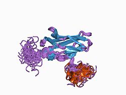
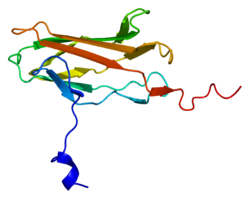
Interactions

RUNX3 has been shown to interact with TLE1. [15]

See also

Related Research Articles

Carcinogenesis, also called oncogenesis or tumorigenesis, is the formation of a cancer, whereby normal cells are transformed into cancer cells. The process is characterized by changes at the cellular, genetic, and epigenetic levels and abnormal cell division. Cell division is a physiological process that occurs in almost all tissues and under a variety of circumstances. Normally, the balance between proliferation and programmed cell death, in the form of apoptosis, is maintained to ensure the integrity of tissues and organs. According to the prevailing accepted theory of carcinogenesis, the somatic mutation theory, mutations in DNA and epimutations that lead to cancer disrupt these orderly processes by interfering with the programming regulating the processes, upsetting the normal balance between proliferation and cell death. This results in uncontrolled cell division and the evolution of those cells by natural selection in the body. Only certain mutations lead to cancer whereas the majority of mutations do not.

<span class="mw-page-title-main">Von Hippel–Lindau tumor suppressor</span> Mammalian protein found in Homo sapiens

The Von Hippel–Lindau tumor suppressor also known as pVHL is a protein that, in humans, is encoded by the VHL gene. Mutations of the VHL gene are associated with Von Hippel–Lindau disease, which is characterized by hemangioblastomas of the brain, spinal cord and retina. It is also associated with kidney and pancreatic lesions.

<span class="mw-page-title-main">RUNX2</span> Protein-coding gene in humans

Runt-related transcription factor 2 (RUNX2) also known as core-binding factor subunit alpha-1 (CBF-alpha-1) is a protein that in humans is encoded by the RUNX2 gene. RUNX2 is a key transcription factor associated with osteoblast differentiation.

<span class="mw-page-title-main">Transcription factor Jun</span> Mammalian protein found in Homo sapiens

Transcription factor Jun is a protein that in humans is encoded by the JUN gene. c-Jun, in combination with protein c-Fos, forms the AP-1 early response transcription factor. It was first identified as the Fos-binding protein p39 and only later rediscovered as the product of the JUN gene. c-jun was the first oncogenic transcription factor discovered. The proto-oncogene c-Jun is the cellular homolog of the viral oncoprotein v-jun. The viral homolog v-jun was discovered in avian sarcoma virus 17 and was named for ju-nana, the Japanese word for 17. The human JUN encodes a protein that is highly similar to the viral protein, which interacts directly with specific target DNA sequences to regulate gene expression. This gene is intronless and is mapped to 1p32-p31, a chromosomal region involved in both translocations and deletions in human malignancies.

<span class="mw-page-title-main">Alpha-enolase</span> Protein-coding gene in Homo sapiens

Enolase 1 (ENO1), more commonly known as alpha-enolase, is a glycolytic enzyme expressed in most tissues, one of the isozymes of enolase. Each isoenzyme is a homodimer composed of 2 alpha, 2 gamma, or 2 beta subunits, and functions as a glycolytic enzyme. Alpha-enolase, in addition, functions as a structural lens protein (tau-crystallin) in the monomeric form. Alternative splicing of this gene results in a shorter isoform that has been shown to bind to the c-myc promoter and function as a tumor suppressor. Several pseudogenes have been identified, including one on the long arm of chromosome 1. Alpha-enolase has also been identified as an autoantigen in Hashimoto encephalopathy.

<span class="mw-page-title-main">RUNX1</span> Protein-coding gene in humans

Runt-related transcription factor 1 (RUNX1) also known as acute myeloid leukemia 1 protein (AML1) or core-binding factor subunit alpha-2 (CBFA2) is a protein that in humans is encoded by the RUNX1 gene.

<span class="mw-page-title-main">KLF4</span> Protein-coding gene in the species Homo sapiens

Kruppel-like factor 4 is a member of the KLF family of zinc finger transcription factors, which belongs to the relatively large family of SP1-like transcription factors. KLF4 is involved in the regulation of proliferation, differentiation, apoptosis and somatic cell reprogramming. Evidence also suggests that KLF4 is a tumor suppressor in certain cancers, including colorectal cancer. It has three C2H2-zinc fingers at its carboxyl terminus that are closely related to another KLF, KLF2. It has two nuclear localization sequences that signals it to localize to the nucleus. In embryonic stem cells (ESCs), KLF4 has been demonstrated to be a good indicator of stem-like capacity. It is suggested that the same is true in mesenchymal stem cells (MSCs).

<span class="mw-page-title-main">CBFB</span> Protein-coding gene in the species Homo sapiens

Core-binding factor subunit beta is a protein that in humans is encoded by the CBFB gene.

<span class="mw-page-title-main">RUNX1T1</span> Protein-coding gene in humans

Protein CBFA2T1 is a protein that in humans is encoded by the RUNX1T1 gene.

<span class="mw-page-title-main">Secreted frizzled-related protein 1</span> Protein-coding gene in the species Homo sapiens

Secreted frizzled-related protein 1, also known as SFRP1, is a protein which in humans is encoded by the SFRP1 gene.

<span class="mw-page-title-main">TLE1</span> Protein-coding gene in the species Homo sapiens

Transducin-like enhancer protein 1 is a protein that in humans is encoded by the TLE1 gene.

<span class="mw-page-title-main">HOXA5</span> Protein-coding gene in humans

Homeobox protein Hox-A5 is a protein that in humans is encoded by the HOXA5 gene.

<span class="mw-page-title-main">XAF1</span> Protein-coding gene in the species Homo sapiens

XIAP-associated factor 1 is a protein that in humans is encoded by the XAF1 gene.

<span class="mw-page-title-main">ID4</span> Protein-coding gene in humans

ID4 is a protein coding gene. In humans, it encodes for the protein known as DNA-binding protein inhibitor ID-4. This protein is known to be involved in the regulation of many cellular processes during both prenatal development and tumorigenesis. This is inclusive of embryonic cellular growth, senescence, cellular differentiation, apoptosis, and as an oncogene in angiogenesis.

<span class="mw-page-title-main">ATOH1</span> Protein-coding gene in the species Homo sapiens

Protein atonal homolog 1 is a protein that in humans is encoded by the ATOH1 gene.

<span class="mw-page-title-main">GRLF1</span> Protein-coding gene in the species Homo sapiens

Glucocorticoid receptor DNA-binding factor 1 is a protein that in humans is encoded by the GRLF1 gene.

<span class="mw-page-title-main">CBFA2T3</span> Protein-coding gene in the species Homo sapiens

Protein CBFA2T3 is a protein that in humans is encoded by the CBFA2T3 gene.

<span class="mw-page-title-main">FOXJ1</span> Protein-coding gene in the species Homo sapiens

Forkhead box protein J1 is a protein that in humans is encoded by the FOXJ1 gene. It is a member of the Forkhead/winged helix (FOX) family of transcription factors that is involved in ciliogenesis. FOXJ1 is expressed in ciliated cells of the lung, choroid plexus, reproductive tract, embryonic kidney and pre-somite embryo stage.

<span class="mw-page-title-main">Cancer epigenetics</span> Field of study in cancer research

Cancer epigenetics is the study of epigenetic modifications to the DNA of cancer cells that do not involve a change in the nucleotide sequence, but instead involve a change in the way the genetic code is expressed. Epigenetic mechanisms are necessary to maintain normal sequences of tissue specific gene expression and are crucial for normal development. They may be just as important, if not even more important, than genetic mutations in a cell's transformation to cancer. The disturbance of epigenetic processes in cancers, can lead to a loss of expression of genes that occurs about 10 times more frequently by transcription silencing than by mutations. As Vogelstein et al. points out, in a colorectal cancer there are usually about 3 to 6 driver mutations and 33 to 66 hitchhiker or passenger mutations. However, in colon tumors compared to adjacent normal-appearing colonic mucosa, there are about 600 to 800 heavily methylated CpG islands in the promoters of genes in the tumors while these CpG islands are not methylated in the adjacent mucosa. Manipulation of epigenetic alterations holds great promise for cancer prevention, detection, and therapy. In different types of cancer, a variety of epigenetic mechanisms can be perturbed, such as the silencing of tumor suppressor genes and activation of oncogenes by altered CpG island methylation patterns, histone modifications, and dysregulation of DNA binding proteins. There are several medications which have epigenetic impact, that are now used in a number of these diseases.

Generally, in progression to cancer, hundreds of genes are silenced or activated. Although silencing of some genes in cancers occurs by mutation, a large proportion of carcinogenic gene silencing is a result of altered DNA methylation. DNA methylation causing silencing in cancer typically occurs at multiple CpG sites in the CpG islands that are present in the promoters of protein coding genes.

References

  1. 1 2 3 GRCh38: Ensembl release 89: ENSG00000020633 - Ensembl, May 2017
  2. 1 2 3 GRCm38: Ensembl release 89: ENSMUSG00000070691 - Ensembl, May 2017
  3. "Human PubMed Reference:". National Center for Biotechnology Information, U.S. National Library of Medicine.
  4. "Mouse PubMed Reference:". National Center for Biotechnology Information, U.S. National Library of Medicine.
  5. Levanon D, Negreanu V, Bernstein Y, Bar-Am I, Avivi L, Groner Y (Sep 1994). "AML1, AML2, and AML3, the human members of the runt domain gene-family: cDNA structure, expression, and chromosomal localization". Genomics. 23 (2): 425–32. doi:10.1006/geno.1994.1519. PMID   7835892.
  6. Levanon D, Eisenstein M, Groner Y (Apr 1998). "Site-directed mutagenesis supports a three-dimensional model of the runt domain". Journal of Molecular Biology. 277 (3): 509–12. doi:10.1006/jmbi.1998.1633. PMID   9533875. S2CID   13139512.
  7. "Entrez Gene: RUNX3 runt-related transcription factor 3".
  8. Hoek KS, Schlegel NC, Eichhoff OM, Widmer DS, Praetorius C, Einarsson SO, Valgeirsdottir S, Bergsteinsdottir K, Schepsky A, Dummer R, Steingrimsson E (Dec 2008). "Novel MITF targets identified using a two-step DNA microarray strategy". Pigment Cell & Melanoma Research. 21 (6): 665–76. doi: 10.1111/j.1755-148X.2008.00505.x . PMID   19067971.
  9. Lee Y, Lee J, Jang J, Chi X, Kim J, Li Y, Kim M, Kim D, Choi B, Kim E, Chung J, Lee O, Lee Y, Suh J, Chuang LS (2013-11-11). "Runx3 inactivation is a crucial early event in the development of lung adenocarcinoma". Cancer Cell. 24 (5): 603–616. doi: 10.1016/j.ccr.2013.10.003 . ISSN   1878-3686. PMID   24229708.
  10. 1 2 3 Lee J, Kim D, Jang J, Park T, Song S, Lee Y, Chi X, Park IY, Hyun J, Ito Y, Bae S (2019-04-23). "RUNX3 regulates cell cycle-dependent chromatin dynamics by functioning as a pioneer factor of the restriction-point". Nature Communications. 10 (1): 1897. Bibcode:2019NatCo..10.1897L. doi: 10.1038/s41467-019-09810-w . ISSN   2041-1723. PMC   6479060 . PMID   31015486.
  11. Li QL, Ito K, Sakakura C, Fukamachi H, Inoue Ki, Chi XZ, Lee KY, Nomura S, Lee CW, Han SB, Kim HM, Kim WJ, Yamamoto H, Yamashita N, Yano T, Ikeda T, Itohara S, Inazawa J, Abe T, Hagiwara A, Yamagishi H, Ooe A, Kaneda A, Sugimura T, Ushijima T, Bae SC, Ito Y (Apr 2002). "Causal relationship between the loss of RUNX3 expression and gastric cancer". Cell. 109 (1): 113–24. doi: 10.1016/S0092-8674(02)00690-6 . PMID   11955451. S2CID   11362226.
  12. Levanon D, Bernstein Y, Negreanu V, Bone KR, Pozner A, Eilam R, Lotem J, Brenner O, Groner Y (Oct 2011). "Absence of Runx3 expression in normal gastrointestinal epithelium calls into question its tumour suppressor function". EMBO Mol Med. 3 (10): 593–604. doi:10.1002/emmm.201100168. PMC   3258485 . PMID   21786422.
  13. Maiväli Ü (2015). Interpreting Biomedical Science. Academic Press. pp. 44–45. ISBN   9780124186897.
  14. Ito K, Inoue K, Bae S, Ito Y (2009-03-12). "Runx3 expression in gastrointestinal tract epithelium: resolving the controversy". Oncogene. 28 (10): 1379–1384. doi:10.1038/onc.2008.496. ISSN   1476-5594. PMID   19169278.
  15. Levanon D, Goldstein RE, Bernstein Y, Tang H, Goldenberg D, Stifani S, Paroush Z, Groner Y (Sep 1998). "Transcriptional repression by AML1 and LEF-1 is mediated by the TLE/Groucho corepressors". Proceedings of the National Academy of Sciences of the United States of America. 95 (20): 11590–5. Bibcode:1998PNAS...9511590L. doi: 10.1073/pnas.95.20.11590 . PMC   21685 . PMID   9751710.

Further reading

This article incorporates text from the United States National Library of Medicine, which is in the public domain.