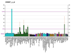
Achaete-scute homolog 1 is a protein that in humans is encoded by the ASCL1 gene. [5] [6] Because it was discovered subsequent to studies on its homolog in Drosophila, the Achaete-scute complex, it was originally named MASH-1 for mammalian achaete scute homolog-1. [7]
This gene encodes a member of the basic helix-loop-helix (BHLH) family of transcription factors. The protein activates transcription by binding to the E box (5'-CANNTG-3'). Dimerization with other BHLH proteins is required for efficient DNA binding. This protein plays a role in the neuronal commitment and differentiation and in the generation of olfactory and autonomic neurons. It is highly expressed in medullary thyroid cancer and small cell lung cancer and may be a useful marker for these cancers. The presence of a CAG repeat in the gene suggests that it may also play a role in tumor formation. [6]
Development of the vertebrate nervous system begins when the neural tube forms in the early embryo. The neural tube eventually gives rise to the entire nervous system, but first neuroblasts must differentiate from the neuroepithelium of the tube. The neuroblasts are the cells that undergo mitotic division and produce neurons. [7] Asc is central to the differentiation of the neuroblasts and the lateral inhibition mechanism which inherently creates a safety net in the event of damage or death in these incredibly important cells. [7]
Differentiation of the neuroblast begins when the cells of the neural tube express Asc and thus upregulate the expression of Delta, a protein essential to the lateral inhibition pathway of neuronal commitment. [7] Delta can diffuse to neighboring cells and bind to the Notch receptor, a large transmembrane protein which upon activation undergoes proteolytic cleavage to release the intracellular domain (Notch-ICD). [7] The Notch-ICD is then free to travel to the nucleus and form a complex with Suppressor of Hairless (SuH) and Mastermind. [7] This complex acts as transcription regulator of Asc and accomplishes two important tasks. First, it prevents the expression of factors required for differentiation of the cell into a neuroblast. [7] Secondly, it inhibits the neighboring cell's production of Delta. [7] Therefore, the future neuroblast will be the cell that has the greatest Asc activation in the vicinity and consequently the greatest Delta production that will inhibit the differentiation of neighboring cells. The select group of neuroblasts that then differentiate in the neural tube are thus replaceable because the neuroblast's ability to suppress differentiation of neighboring cells depends on its own ability to produce Asc. [7] This process of neuroblast differentiation via Asc is common to all animals. [7] Although this mechanism was initially studied in Drosophila, homologs to all proteins in the pathway have been found in vertebrates that have the same bHLH structure. [7]
In addition to its important role in neuroblast formation, Asc also functions to mediate autonomic nervous system (ANS) formation. [8] Asc was initially suspected to play a role in the ANS when ASCL1 was found expressed in cells surrounding the dorsal aorta, the adrenal glands and in the developing sympathetic chain during a specific stage of development. [8] Subsequent studies of mice genetically altered to be MASH-1 deficient revealed defective development of both sympathetic and parasympathetic ganglia, the two constituents of the ANS. [8]
ASCL1 has been shown to interact with Myocyte-specific enhancer factor 2A. [9]
In vertebrates, a neuroblast or primitive nerve cell is a postmitotic cell that does not divide further, and which will develop into a neuron after a migration phase. In invertebrates such as Drosophila, neuroblasts are neural progenitor cells which divide asymmetrically to produce a neuroblast, and a daughter cell of varying potency depending on the type of neuroblast. Vertebrate neuroblasts differentiate from radial glial cells and are committed to becoming neurons. Neural stem cells, which only divide symmetrically to produce more neural stem cells, transition gradually into radial glial cells. Radial glial cells, also called radial glial progenitor cells, divide asymmetrically to produce a neuroblast and another radial glial cell that will re-enter the cell cycle.
The Notch signaling pathway is a highly conserved cell signaling system present in most animals. Mammals possess four different notch receptors, referred to as NOTCH1, NOTCH2, NOTCH3, and NOTCH4. The notch receptor is a single-pass transmembrane receptor protein. It is a hetero-oligomer composed of a large extracellular portion, which associates in a calcium-dependent, non-covalent interaction with a smaller piece of the notch protein composed of a short extracellular region, a single transmembrane-pass, and a small intracellular region.
An asymmetric cell division produces two daughter cells with different cellular fates. This is in contrast to symmetric cell divisions which give rise to daughter cells of equivalent fates. Notably, stem cells divide asymmetrically to give rise to two distinct daughter cells: one copy of the original stem cell as well as a second daughter programmed to differentiate into a non-stem cell fate.
Neurogenic locus notch homolog protein 3(Notch 3) is a protein that in humans is encoded by the NOTCH3 gene.
The gene extramachrochaetae (emc) is a Drosophila melanogaster gene that codes for the Emc protein, which has a wide variety of developmental roles. It was named, as is common for Drosophila genes, after the phenotypic change caused by a mutation in the gene (macrochaetae are the longer bristles on Drosophila).
Neurogenic locus notch homolog protein 1(Notch 1) is a protein encoded in humans by the NOTCH1 gene. Notch 1 is a single-pass transmembrane receptor.
Enhancer of zeste homolog 2 (EZH2) is a histone-lysine N-methyltransferase enzyme encoded by EZH2 gene, that participates in histone methylation and, ultimately, transcriptional repression. EZH2 catalyzes the addition of methyl groups to histone H3 at lysine 27, by using the cofactor S-adenosyl-L-methionine. Methylation activity of EZH2 facilitates heterochromatin formation thereby silences gene function. Remodeling of chromosomal heterochromatin by EZH2 is also required during cell mitosis.
Neurogenic locus notch homolog 4(Notch 4) is a protein that in humans is encoded by the NOTCH4 gene located on chromosome 6.
Protein numb homolog is a protein that in humans is encoded by the NUMB gene. The protein encoded by this gene plays a role in the determination of cell fates during development. The encoded protein, whose degradation is induced in a proteasome-dependent manner by MDM2, is a membrane-bound protein that has been shown to associate with EPS15, LNX1, and NOTCH1. Four transcript variants encoding different isoforms have been found for this gene.
Myocyte-specific enhancer factor 2A is a protein that in humans is encoded by the MEF2A gene. MEF2A is a transcription factor in the Mef2 family. In humans it is located on chromosome 15q26. Certain mutations in MEF2A cause an autosomal dominant form of coronary artery disease and myocardial infarction.
Protein BTG2 also known as BTG family member 2 or NGF-inducible anti-proliferative protein PC3 or NGF-inducible protein TIS21, is a protein that in humans is encoded by the BTG2 gene and in other mammals by the homologous Btg2 gene. This protein controls cell cycle progression and proneural genes expression by acting as a transcription coregulator that enhances or inhibits the activity of transcription factors.
Transducin-like enhancer protein 1 is a protein that in humans is encoded by the TLE1 gene.
Transcription factor HES1 is a protein that is encoded by the Hes1 gene, and is the mammalian homolog of the hairy gene in Drosophila. HES1 is one of the seven members of the Hes gene family (HES1-7). Hes genes code nuclear proteins that suppress transcription.
Single-minded homolog 2 is a protein that in humans is encoded by the SIM2 gene. It plays a major role in the development of the central nervous system midline as well as the construction of the face and head.
Protein atonal homolog 1 is a protein that in humans is encoded by the ATOH1 gene.
Neurogenins are a family of bHLH transcription factors involved in specifying neuronal differentiation. It is one of many gene families related to the atonal gene in Drosophila. Other positive regulators of neuronal differentiation also expressed during early neural development include NeuroD and ASCL1.
Achaete-scute complex homolog 2 (Drosophila), also known as ASCL2, is an imprinted human gene.
Ganglion mother cells (GMCs) are cells involved in neurogenesis, in non-mammals, that divide only once to give rise to two neurons, or one neuron and one glial cell or two glial cells, and are present only in the central nervous system. They are also responsible for transcription factor expression. While each ganglion mother cell necessarily gives rise to two neurons, a neuroblast can asymmetrically divide multiple times. GMCs are the progeny of type I neuroblasts. Neuroblasts asymmetrically divide during embryogenesis to create GMCs. GMCs are only present in certain species and only during the embryonic and larval stages of life. Recent research has shown that there is an intermediate stage between a GMC and two neurons. The GMC forms two ganglion cells which then develop into neurons or glial cells. Embryonic neurogenesis has been extensively studied in Drosophila melanogaster embryos and larvae.
The achaete-scute complex (AS-C) is a group of four genes in the fruit fly Drosophila melanogaster. These genes encode basic helix-loop-helix transcription factors that have been best studied in their regulation of nervous system development. Because of their role in specifying neuroblast fate, the genes of the AS-C are called proneural genes. However, the AS-C has non-proneural functions, such as specifying muscle and gut progenitors. Homologues of AS-C in other animals, including humans and other vertebrates, have similar functions.
Proneural genes encode transcription factors of the basic helix-loop-helix (bHLH) class which are responsible for the development of neuroectodermal progenitor cells. Proneural genes have multiple functions in neural development. They integrate positional information and contribute to the specification of progenitor-cell identity. From the same ectodermal cell types, neural or epidermal cells can develop based on interactions between proneural and neurogenic genes. Neurogenic genes are so called because loss of function mutants show an increase number of developed neural precursors. On the other hand, proneural genes mutants fail to develop neural precursor cells.
This article incorporates text from the United States National Library of Medicine, which is in the public domain.